+ Open data
- Basic information
| Entry | Database: EMDB / ID: EMD-12336 | ||||||||||||
|---|---|---|---|---|---|---|---|---|---|---|---|---|---|
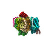
| Title | Structure of PSII-I (PSII with Psb27, Psb28, and Psb34) | ||||||||||||
 Map data Map data | CryoEM Map of PSII-I (PSII-Psb28-Psb34-Psb27) | ||||||||||||
 Sample Sample |
| ||||||||||||
 Keywords Keywords | Membrane Protein Biogenesis Assembly Factors Photosystem II / PHOTOSYNTHESIS | ||||||||||||
| Function / homology |  Function and homology information Function and homology informationphotosystem II repair / thylakoid lumen / thylakoid / photosystem II oxygen evolving complex / photosystem II assembly / oxygen evolving activity / photosystem II stabilization / photosystem II reaction center / photosystem II / oxidoreductase activity, acting on diphenols and related substances as donors, oxygen as acceptor ...photosystem II repair / thylakoid lumen / thylakoid / photosystem II oxygen evolving complex / photosystem II assembly / oxygen evolving activity / photosystem II stabilization / photosystem II reaction center / photosystem II / oxidoreductase activity, acting on diphenols and related substances as donors, oxygen as acceptor / photosynthetic electron transport chain / photosystem II / response to herbicide / : / plasma membrane-derived thylakoid membrane / chlorophyll binding / photosynthetic electron transport in photosystem II / phosphate ion binding / photosynthesis, light reaction / photosynthesis / electron transfer activity / protein stabilization / iron ion binding / heme binding / metal ion binding Similarity search - Function | ||||||||||||
| Biological species |   Thermosynechococcus elongatus BP-1 (bacteria) Thermosynechococcus elongatus BP-1 (bacteria) | ||||||||||||
| Method | single particle reconstruction / cryo EM / Resolution: 2.72 Å | ||||||||||||
 Authors Authors | Zabret J / Bohn S | ||||||||||||
| Funding support |  Germany, 3 items Germany, 3 items
| ||||||||||||
 Citation Citation |  Journal: Nat Plants / Year: 2021 Journal: Nat Plants / Year: 2021Title: Structural insights into photosystem II assembly. Authors: Jure Zabret / Stefan Bohn / Sandra K Schuller / Oliver Arnolds / Madeline Möller / Jakob Meier-Credo / Pasqual Liauw / Aaron Chan / Emad Tajkhorshid / Julian D Langer / Raphael Stoll / Anja ...Authors: Jure Zabret / Stefan Bohn / Sandra K Schuller / Oliver Arnolds / Madeline Möller / Jakob Meier-Credo / Pasqual Liauw / Aaron Chan / Emad Tajkhorshid / Julian D Langer / Raphael Stoll / Anja Krieger-Liszkay / Benjamin D Engel / Till Rudack / Jan M Schuller / Marc M Nowaczyk /    Abstract: Biogenesis of photosystem II (PSII), nature's water-splitting catalyst, is assisted by auxiliary proteins that form transient complexes with PSII components to facilitate stepwise assembly events. ...Biogenesis of photosystem II (PSII), nature's water-splitting catalyst, is assisted by auxiliary proteins that form transient complexes with PSII components to facilitate stepwise assembly events. Using cryo-electron microscopy, we solved the structure of such a PSII assembly intermediate from Thermosynechococcus elongatus at 2.94 Å resolution. It contains three assembly factors (Psb27, Psb28 and Psb34) and provides detailed insights into their molecular function. Binding of Psb28 induces large conformational changes at the PSII acceptor side, which distort the binding pocket of the mobile quinone (Q) and replace the bicarbonate ligand of non-haem iron with glutamate, a structural motif found in reaction centres of non-oxygenic photosynthetic bacteria. These results reveal mechanisms that protect PSII from damage during biogenesis until water splitting is activated. Our structure further demonstrates how the PSII active site is prepared for the incorporation of the MnCaO cluster, which performs the unique water-splitting reaction. | ||||||||||||
| History |
|
- Structure visualization
| Movie |
 Movie viewer Movie viewer |
|---|---|
| Structure viewer | EM map:  SurfView SurfView Molmil Molmil Jmol/JSmol Jmol/JSmol |
| Supplemental images |
- Downloads & links
-EMDB archive
| Map data |  emd_12336.map.gz emd_12336.map.gz | 51.8 MB |  EMDB map data format EMDB map data format | |
|---|---|---|---|---|
| Header (meta data) |  emd-12336-v30.xml emd-12336-v30.xml emd-12336.xml emd-12336.xml | 30.6 KB 30.6 KB | Display Display |  EMDB header EMDB header |
| Images |  emd_12336.png emd_12336.png | 62.7 KB | ||
| Filedesc metadata |  emd-12336.cif.gz emd-12336.cif.gz | 7.8 KB | ||
| Archive directory |  http://ftp.pdbj.org/pub/emdb/structures/EMD-12336 http://ftp.pdbj.org/pub/emdb/structures/EMD-12336 ftp://ftp.pdbj.org/pub/emdb/structures/EMD-12336 ftp://ftp.pdbj.org/pub/emdb/structures/EMD-12336 | HTTPS FTP |
-Validation report
| Summary document |  emd_12336_validation.pdf.gz emd_12336_validation.pdf.gz | 535.2 KB | Display |  EMDB validaton report EMDB validaton report |
|---|---|---|---|---|
| Full document |  emd_12336_full_validation.pdf.gz emd_12336_full_validation.pdf.gz | 534.8 KB | Display | |
| Data in XML |  emd_12336_validation.xml.gz emd_12336_validation.xml.gz | 6.1 KB | Display | |
| Data in CIF |  emd_12336_validation.cif.gz emd_12336_validation.cif.gz | 7 KB | Display | |
| Arichive directory |  https://ftp.pdbj.org/pub/emdb/validation_reports/EMD-12336 https://ftp.pdbj.org/pub/emdb/validation_reports/EMD-12336 ftp://ftp.pdbj.org/pub/emdb/validation_reports/EMD-12336 ftp://ftp.pdbj.org/pub/emdb/validation_reports/EMD-12336 | HTTPS FTP |
-Related structure data
| Related structure data |  7nhpMC  7nhoC  7nhqC M: atomic model generated by this map C: citing same article ( |
|---|---|
| Similar structure data |
- Links
Links
| EMDB pages |  EMDB (EBI/PDBe) / EMDB (EBI/PDBe) /  EMDataResource EMDataResource |
|---|---|
| Related items in Molecule of the Month |
- Map
Map
| File |  Download / File: emd_12336.map.gz / Format: CCP4 / Size: 67 MB / Type: IMAGE STORED AS FLOATING POINT NUMBER (4 BYTES) Download / File: emd_12336.map.gz / Format: CCP4 / Size: 67 MB / Type: IMAGE STORED AS FLOATING POINT NUMBER (4 BYTES) | ||||||||||||||||||||||||||||||||||||||||||||||||||||||||||||
|---|---|---|---|---|---|---|---|---|---|---|---|---|---|---|---|---|---|---|---|---|---|---|---|---|---|---|---|---|---|---|---|---|---|---|---|---|---|---|---|---|---|---|---|---|---|---|---|---|---|---|---|---|---|---|---|---|---|---|---|---|---|
| Annotation | CryoEM Map of PSII-I (PSII-Psb28-Psb34-Psb27) | ||||||||||||||||||||||||||||||||||||||||||||||||||||||||||||
| Projections & slices | Image control
Images are generated by Spider. | ||||||||||||||||||||||||||||||||||||||||||||||||||||||||||||
| Voxel size | X=Y=Z: 1.09 Å | ||||||||||||||||||||||||||||||||||||||||||||||||||||||||||||
| Density |
| ||||||||||||||||||||||||||||||||||||||||||||||||||||||||||||
| Symmetry | Space group: 1 | ||||||||||||||||||||||||||||||||||||||||||||||||||||||||||||
| Details | EMDB XML:
CCP4 map header:
| ||||||||||||||||||||||||||||||||||||||||||||||||||||||||||||
-Supplemental data
- Sample components
+Entire : PsII-I
+Supramolecule #1: PsII-I
+Macromolecule #1: Photosystem II protein D1 1
+Macromolecule #2: Photosystem II CP47 reaction center protein
+Macromolecule #3: Photosystem II CP43 reaction center protein
+Macromolecule #4: Photosystem II D2 protein
+Macromolecule #5: Cytochrome b559 subunit alpha
+Macromolecule #6: Cytochrome b559 subunit beta
+Macromolecule #7: Photosystem II reaction center protein H
+Macromolecule #8: Photosystem II reaction center protein I
+Macromolecule #9: Photosystem II reaction center protein K
+Macromolecule #10: Photosystem II reaction center protein L
+Macromolecule #11: Photosystem II reaction center protein M
+Macromolecule #12: Photosystem II reaction center protein T
+Macromolecule #13: Photosystem II reaction center X protein
+Macromolecule #14: Photosystem II reaction center protein Ycf12
+Macromolecule #15: Photosystem II reaction center protein Z
+Macromolecule #16: Photosystem II lipoprotein Psb27
+Macromolecule #17: Photosystem II reaction center Psb28 protein
+Macromolecule #18: Tsl0063 protein
+Macromolecule #19: MANGANESE (II) ION
+Macromolecule #20: CHLORIDE ION
+Macromolecule #21: PHEOPHYTIN A
+Macromolecule #22: CHLOROPHYLL A
+Macromolecule #23: BETA-CAROTENE
+Macromolecule #24: 1,2-DIPALMITOYL-PHOSPHATIDYL-GLYCEROLE
+Macromolecule #25: 1,2-DISTEAROYL-MONOGALACTOSYL-DIGLYCERIDE
+Macromolecule #26: FE (III) ION
+Macromolecule #27: 2,3-DIMETHYL-5-(3,7,11,15,19,23,27,31,35-NONAMETHYL-2,6,10,14,18,...
+Macromolecule #28: PROTOPORPHYRIN IX CONTAINING FE
-Experimental details
-Structure determination
| Method | cryo EM |
|---|---|
 Processing Processing | single particle reconstruction |
| Aggregation state | particle |
- Sample preparation
| Buffer | pH: 6.5 |
|---|---|
| Vitrification | Cryogen name: ETHANE-PROPANE |
- Electron microscopy
| Microscope | FEI TITAN KRIOS |
|---|---|
| Image recording | Film or detector model: GATAN K3 (6k x 4k) / Average electron dose: 55.0 e/Å2 |
| Electron beam | Acceleration voltage: 300 kV / Electron source:  FIELD EMISSION GUN FIELD EMISSION GUN |
| Electron optics | Illumination mode: FLOOD BEAM / Imaging mode: BRIGHT FIELD |
| Experimental equipment |  Model: Titan Krios / Image courtesy: FEI Company |
- Image processing
| Startup model | Type of model: NONE |
|---|---|
| Final reconstruction | Resolution.type: BY AUTHOR / Resolution: 2.72 Å / Resolution method: FSC 0.143 CUT-OFF / Number images used: 57268 |
| Initial angle assignment | Type: PROJECTION MATCHING |
| Final angle assignment | Type: PROJECTION MATCHING |
 Movie
Movie Controller







 Z (Sec.)
 Y (Row.)
 X (Col.)


