 Movie
Movie Controller


+ Open data

- Basic information
| Entry | Database: EMDB / ID: EMD-1232 | |||||||||
|---|---|---|---|---|---|---|---|---|---|---|
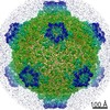
| Title | Structure of the Sec13-31 COPII coat cage. | |||||||||
 Map data Map data | This is a 30 angstrom resolution map of the self-assembing Sec13/31 cage. | |||||||||
 Sample Sample |
| |||||||||
| Function / homology | COPII vesicle coat / intracellular protein transport / WD40 repeat Function and homology information Function and homology information | |||||||||
| Biological species |  Homo sapiens (human) Homo sapiens (human) | |||||||||
| Method | single particle reconstruction / cryo EM / Resolution: 30.0 Å | |||||||||
 Authors Authors | Stagg SM / Gurkan C / Fowler DM / LaPointe P / Foss TR / Potter CS / Carragher B / Balch WE | |||||||||
 Citation Citation | Journal: Nature / Year: 2006 Title: Structure of the Sec13/31 COPII coat cage. Authors: Scott M Stagg / Cemal Gürkan / Douglas M Fowler / Paul LaPointe / Ted R Foss / Clinton S Potter / Bridget Carragher / William E Balch /  Abstract: Endomembranes of eukaryotic cells are dynamic structures that are in continuous communication through the activity of specialized cellular machineries, such as the coat protein complex II (COPII), ...Endomembranes of eukaryotic cells are dynamic structures that are in continuous communication through the activity of specialized cellular machineries, such as the coat protein complex II (COPII), which mediates cargo export from the endoplasmic reticulum (ER). COPII consists of the Sar1 GTPase, Sec23 and Sec24 (Sec23/24), where Sec23 is a Sar1-specific GTPase-activating protein and Sec24 functions in cargo selection, and Sec13 and Sec31 (Sec13/31), which has a structural role. Whereas recent results have shown that Sec23/24 and Sec13/31 can self-assemble to form COPII cage-like particles, we now show that Sec13/31 can self-assemble to form minimal cages in the absence of Sec23/24. We present a three-dimensional reconstruction of these Sec13/31 cages at 30 A resolution using cryo-electron microscopy and single particle analysis. These results reveal a novel cuboctahedron geometry with the potential to form a flexible lattice and to generate a diverse range of containers. Our data are consistent with a model for COPII coat complex assembly in which Sec23/24 has a non-structural role as a multivalent ligand localizing the self-assembly of Sec13/31 to form a cage lattice driving ER cargo export. | |||||||||
| History |
|
- Structure visualization
| Movie |
Movie viewer |
|---|---|
| Structure viewer | EM map: SurfViewMolmilJmol/JSmol |
| Supplemental images |

- Downloads & links
-EMDB archive
| Map data | emd_1232.map.gz | 21 MB | EMDB map data format | |
|---|---|---|---|---|
| Header (meta data) | emd-1232-v30.xmlemd-1232.xml | 10.8 KB 10.8 KB | Display Display | EMDB header |
| Images |  1232.gif 1232.gif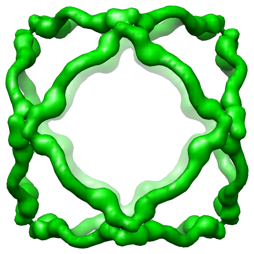 EMD-1232.png EMD-1232.png | 13.2 KB 137.8 KB | ||
| Archive directory |  http://ftp.pdbj.org/pub/emdb/structures/EMD-1232ftp://ftp.pdbj.org/pub/emdb/structures/EMD-1232 http://ftp.pdbj.org/pub/emdb/structures/EMD-1232ftp://ftp.pdbj.org/pub/emdb/structures/EMD-1232 | HTTPS FTP |
-Related structure data
| Similar structure data |
|---|

-Links
| EMDB pages | EMDB (EBI/PDBe) / EMDataResource |
|---|
-Map
| File | Download / File: emd_1232.map.gz / Format: CCP4 / Size: 26.4 MB / Type: IMAGE STORED AS FLOATING POINT NUMBER (4 BYTES) | ||||||||||||||||||||||||||||||||||||||||||||||||||||||||||||||||||||
|---|---|---|---|---|---|---|---|---|---|---|---|---|---|---|---|---|---|---|---|---|---|---|---|---|---|---|---|---|---|---|---|---|---|---|---|---|---|---|---|---|---|---|---|---|---|---|---|---|---|---|---|---|---|---|---|---|---|---|---|---|---|---|---|---|---|---|---|---|---|
| Annotation | This is a 30 angstrom resolution map of the self-assembing Sec13/31 cage. | ||||||||||||||||||||||||||||||||||||||||||||||||||||||||||||||||||||
| Projections & slices | Image control
Images are generated by Spider. | ||||||||||||||||||||||||||||||||||||||||||||||||||||||||||||||||||||
| Voxel size | X=Y=Z: 4.526 Å | ||||||||||||||||||||||||||||||||||||||||||||||||||||||||||||||||||||
| Density |
| ||||||||||||||||||||||||||||||||||||||||||||||||||||||||||||||||||||
| Symmetry | Space group: 1 | ||||||||||||||||||||||||||||||||||||||||||||||||||||||||||||||||||||
| Details | EMDB XML:
CCP4 map header:
| ||||||||||||||||||||||||||||||||||||||||||||||||||||||||||||||||||||
 Z (Sec.)
 Y (Row.)
 X (Col.)


-Supplemental data
- Sample components
-Entire : Oligomeric assembly of Sec13-31
| Entire | Name: Oligomeric assembly of Sec13-31 |
|---|---|
| Components |
|
-Supramolecule #1000: Oligomeric assembly of Sec13-31
| Supramolecule | Name: Oligomeric assembly of Sec13-31 / type: sample / ID: 1000 Oligomeric state: 24-mer of Sec13-31 heterotetramers forming a cuboctahedron Number unique components: 2 |
|---|---|
| Molecular weight | Theoretical: 8.14 MDa |
-Macromolecule #1: Sec13
| Macromolecule | Name: Sec13 / type: protein_or_peptide / ID: 1 / Name.synonym: Sec13 / Details: SEC13R / Recombinant expression: Yes |
|---|---|
| Source (natural) | Organism: Homo sapiens (human) / synonym: Human |
| Molecular weight | Experimental: 33 MDa / Theoretical: 33 MDa |
| Recombinant expression | Organism: Baculovirus infected Tn5 insect cells / Recombinant plasmid: pFastBac |
| Sequence | GO: intracellular protein transport / InterPro: WD40 repeat |
-Macromolecule #2: Sec31
| Macromolecule | Name: Sec31 / type: protein_or_peptide / ID: 2 / Name.synonym: Sec31 / Details: SEC31L1 / Recombinant expression: Yes |
|---|---|
| Source (natural) | Organism: Homo sapiens (human) / synonym: Human |
| Molecular weight | Experimental: 139 MDa / Theoretical: 139 MDa |
| Recombinant expression | Organism: Baculovirus infected Tn5 insect cells / Recombinant plasmid: pFastBac |
| Sequence | GO: COPII vesicle coat / InterPro: WD40 repeat |
-Experimental details
-Structure determination
| Method | cryo EM |
|---|---|
 Processing Processing | single particle reconstruction |
| Aggregation state | particle |
-Sample preparation
| Concentration | 0.9 mg/mL |
|---|---|
| Buffer | pH: 6.5 / Details: 50mM MES pH 6.5, 225mM KOAc, 1mM MgCl2 |
| Grid | Details: Quantifoil 2/2, 400 mesh copper grid |
| Vitrification | Cryogen name: ETHANE / Chamber humidity: 100 % / Chamber temperature: 93 K / Instrument: OTHER Details: Vitrification instrument: FEI Vitrobot. Grid plasma cleaned for 30s with Fischione 1020 plasma cleaner using 75-25 Argon-Oxygen mix. Method: Temperature of chamber was 4 degrees C. 0 seconds drain time. Single blot. 0 mm offset. 4 ul sample applied to grid. Blot for 3.0 seconds before plunging. |
- Electron microscopy
| Microscope | FEI TECNAI 20 |
|---|---|
| Temperature | Average: 94 K |
| Alignment procedure | Legacy - Astigmatism: objective lens astigmatism was corrected at 50,000X magnification |
| Date | Feb 2, 2005 |
| Image recording | Category: CCD / Film or detector model: GATAN ULTRASCAN 4000 (4k x 4k) / Average electron dose: 20 e/Å2 / Bits/pixel: 16 |
| Electron beam | Acceleration voltage: 200 kV / Electron source:  FIELD EMISSION GUN FIELD EMISSION GUN |
| Electron optics | Illumination mode: FLOOD BEAM / Imaging mode: BRIGHT FIELD / Cs: 2 mm / Nominal defocus max: 2.5 µm / Nominal defocus min: 0.8 µm / Nominal magnification: 50000 |
| Sample stage | Specimen holder: Side entry liquid nitrogen-cooled cryo specimen holder. Specimen holder model: GATAN LIQUID NITROGEN |
-Image processing
| Details | The images were acquired using the Leginon automated data aquisition system. The particles were automatically selected using the Selexon package. The CTF was automatically estimated using the ACE package |
|---|---|
| CTF correction | Details: Phase correction for each particle. |
| Final reconstruction | Applied symmetry - Point group: O (octahedral) / Algorithm: OTHER / Resolution.type: BY AUTHOR / Resolution: 30.0 Å / Resolution method: FSC 0.5 CUT-OFF / Software - Name: EMAN / Number images used: 9777 |
| Final two d classification | Number classes: 92 |
