Movie
Movie Controller


+ Open data

- Basic information
| Entry | Database: EMDB / ID: EMD-12297 | |||||||||
|---|---|---|---|---|---|---|---|---|---|---|
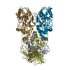
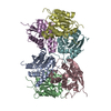
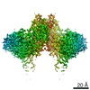
| Title | Ovine rBAT ectodomain homodimer, C2-symmetry | |||||||||
 Map data Map data | Postprocessed map | |||||||||
 Sample Sample |
| |||||||||
 Keywords Keywords | Transporter / Cystinuria / Complex / Ectodomain / TRANSLOCASE | |||||||||
| Function / homology |  Function and homology information Function and homology informationamino acid transport / carbohydrate metabolic process / apical plasma membrane Similarity search - Function | |||||||||
| Biological species |  | |||||||||
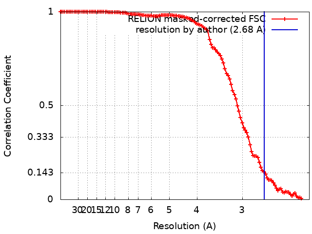
| Method | single particle reconstruction / cryo EM / Resolution: 2.68 Å | |||||||||
 Authors Authors | Lee Y / Kuehlbrandt W | |||||||||
| Funding support |  Germany, 2 items Germany, 2 items
| |||||||||
 Citation Citation | Journal: Nat Commun / Year: 2022 Title: Ca-mediated higher-order assembly of heterodimers in amino acid transport system b biogenesis and cystinuria. Authors: Yongchan Lee / Pattama Wiriyasermkul / Pornparn Kongpracha / Satomi Moriyama / Deryck J Mills / Werner Kühlbrandt / Shushi Nagamori /  Abstract: Cystinuria is a genetic disorder characterized by overexcretion of dibasic amino acids and cystine, causing recurrent kidney stones and kidney failure. Mutations of the regulatory glycoprotein rBAT ...Cystinuria is a genetic disorder characterized by overexcretion of dibasic amino acids and cystine, causing recurrent kidney stones and kidney failure. Mutations of the regulatory glycoprotein rBAT and the amino acid transporter bAT, which constitute system b, are linked to type I and non-type I cystinuria respectively and they exhibit distinct phenotypes due to protein trafficking defects or catalytic inactivation. Here, using electron cryo-microscopy and biochemistry, we discover that Ca mediates higher-order assembly of system b. Ca stabilizes the interface between two rBAT molecules, leading to super-dimerization of bAT-rBAT, which in turn facilitates N-glycan maturation and protein trafficking. A cystinuria mutant T216M and mutations of the Ca site of rBAT cause the loss of higher-order assemblies, resulting in protein trapping at the ER and the loss of function. These results provide the molecular basis of system b biogenesis and type I cystinuria and serve as a guide to develop new therapeutic strategies against it. More broadly, our findings reveal an unprecedented link between transporter oligomeric assembly and protein-trafficking diseases. #1: Journal: Biorxiv / Year: 2021Title: Ca 2+ -mediated higher-order assembly of b 0,+ AT-rBAT is a key step for system b 0,+ biogenesis and cystinuria Authors: Lee Y / Wiriyasermkul P / Moriyama S / Mills DJ / Kuhlbrandt W / Nagamori S | |||||||||
| History |
|
- Structure visualization
| Movie |
Movie viewer |
|---|---|
| Structure viewer | EM map: SurfViewMolmilJmol/JSmol |
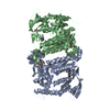
| Supplemental images |
- Downloads & links
-EMDB archive
| Map data | emd_12297.map.gz | 9 MB | EMDB map data format | |
|---|---|---|---|---|
| Header (meta data) | emd-12297-v30.xmlemd-12297.xml | 19.4 KB 19.4 KB | Display Display | EMDB header |
| FSC (resolution estimation) | emd_12297_fsc.xml | 11.3 KB | Display | FSC data file |
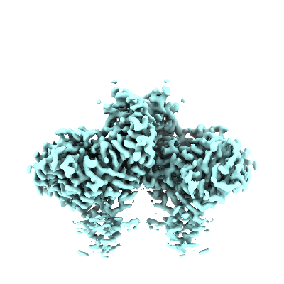
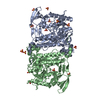
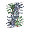
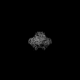
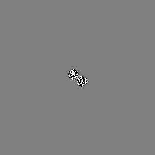
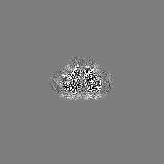
| Images |  emd_12297.png emd_12297.png | 91.1 KB | ||
| Masks | emd_12297_msk_1.map | 125 MB | Mask map | |
| Filedesc metadata | emd-12297.cif.gz | 6.6 KB | ||
| Others | emd_12297_half_map_1.map.gzemd_12297_half_map_2.map.gz | 96.7 MB 96.7 MB | ||
| Archive directory |  http://ftp.pdbj.org/pub/emdb/structures/EMD-12297ftp://ftp.pdbj.org/pub/emdb/structures/EMD-12297 http://ftp.pdbj.org/pub/emdb/structures/EMD-12297ftp://ftp.pdbj.org/pub/emdb/structures/EMD-12297 | HTTPS FTP |
-Related structure data
| Related structure data |  7nf7MC  7nf6C  7nf8C M: atomic model generated by this map C: citing same article ( |
|---|---|
| Similar structure data |


-Links
| EMDB pages | EMDB (EBI/PDBe) / EMDataResource |
|---|---|
| Related items in Molecule of the Month |


-Map
| File | Download / File: emd_12297.map.gz / Format: CCP4 / Size: 125 MB / Type: IMAGE STORED AS FLOATING POINT NUMBER (4 BYTES) | ||||||||||||||||||||||||||||||||||||||||||||||||||||||||||||
|---|---|---|---|---|---|---|---|---|---|---|---|---|---|---|---|---|---|---|---|---|---|---|---|---|---|---|---|---|---|---|---|---|---|---|---|---|---|---|---|---|---|---|---|---|---|---|---|---|---|---|---|---|---|---|---|---|---|---|---|---|---|
| Annotation | Postprocessed map | ||||||||||||||||||||||||||||||||||||||||||||||||||||||||||||
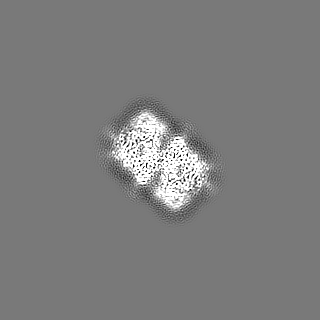
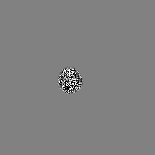
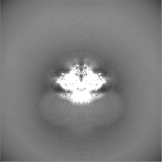
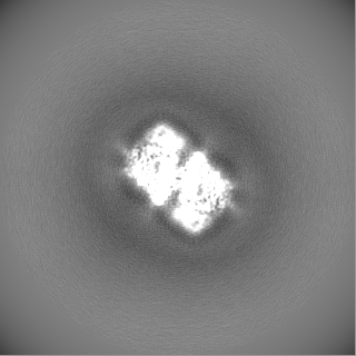
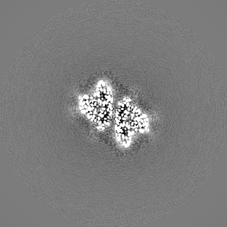
| Projections & slices | Image control
Images are generated by Spider. | ||||||||||||||||||||||||||||||||||||||||||||||||||||||||||||
| Voxel size | X=Y=Z: 1.09856 Å | ||||||||||||||||||||||||||||||||||||||||||||||||||||||||||||
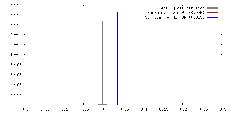
| Density |
| ||||||||||||||||||||||||||||||||||||||||||||||||||||||||||||
| Symmetry | Space group: 1 | ||||||||||||||||||||||||||||||||||||||||||||||||||||||||||||
| Details | EMDB XML:
CCP4 map header:
| ||||||||||||||||||||||||||||||||||||||||||||||||||||||||||||
 Z (Sec.)
 Y (Row.)
 X (Col.)


-Supplemental data
-Mask #1
| File | emd_12297_msk_1.map | ||||||||||||
|---|---|---|---|---|---|---|---|---|---|---|---|---|---|
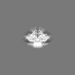
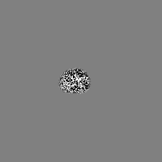
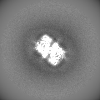
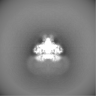
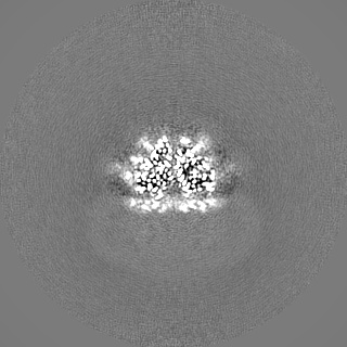
| Projections & Slices |
| ||||||||||||
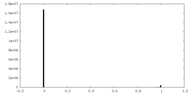
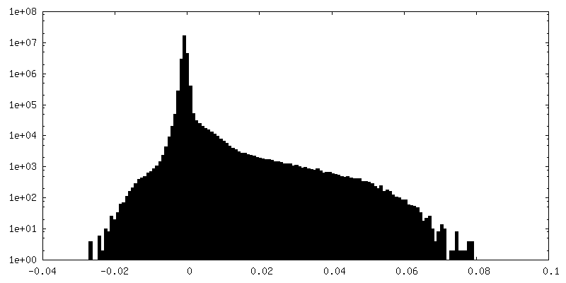
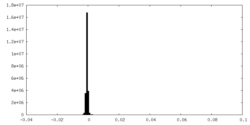
| Density Histograms |


-Half map: Half map 1
| File | emd_12297_half_map_1.map | ||||||||||||
|---|---|---|---|---|---|---|---|---|---|---|---|---|---|
| Annotation | Half map 1 | ||||||||||||
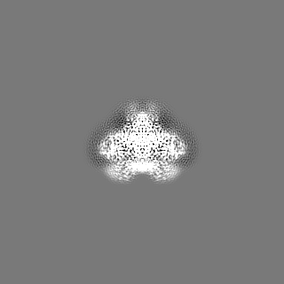
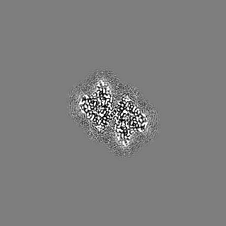
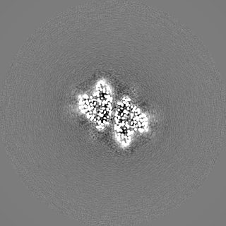
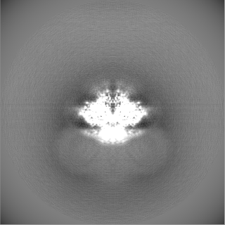
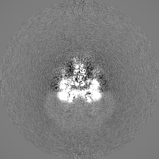
| Projections & Slices |
| ||||||||||||
| Density Histograms |


-Half map: Half map 2
| File | emd_12297_half_map_2.map | ||||||||||||
|---|---|---|---|---|---|---|---|---|---|---|---|---|---|
| Annotation | Half map 2 | ||||||||||||
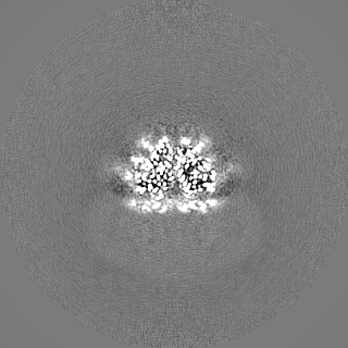
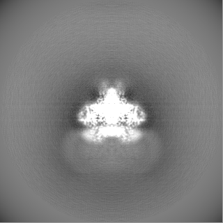
| Projections & Slices |
| ||||||||||||
| Density Histograms |

- Sample components
-Entire : Ovine rBAT ectodomain homodimer
| Entire | Name: Ovine rBAT ectodomain homodimer |
|---|---|
| Components |
|
-Supramolecule #1: Ovine rBAT ectodomain homodimer
| Supramolecule | Name: Ovine rBAT ectodomain homodimer / type: complex / ID: 1 / Parent: 0 / Macromolecule list: #1 |
|---|---|
| Source (natural) | Organism: |
| Molecular weight | Theoretical: 79 kDa/nm |
-Macromolecule #1: neutral and basic amino acid transport protein rBAT
| Macromolecule | Name: neutral and basic amino acid transport protein rBAT / type: protein_or_peptide / ID: 1 / Number of copies: 1 / Enantiomer: LEVO |
|---|---|
| Source (natural) | Organism: |
| Molecular weight | Theoretical: 78.754281 KDa |
| Recombinant expression | Organism:  Homo sapiens (human) Homo sapiens (human) |
| Sequence | String: GSAEDKSKRD SIGLNAKEGQ TNNGFVQNED ILETDLDPSS PAAGPQHNTV DILGPGEPDV KDVRPYAGMP KEVLFQFSGQ ARYRIPREV LFWLTVASVL LLIAATIAII AISPKCLDWW QAGPMYQIYP RSFRDSNKDG DGDLKGIQDK LDYITTLNIK T VWITSFYK ...String: GSAEDKSKRD SIGLNAKEGQ TNNGFVQNED ILETDLDPSS PAAGPQHNTV DILGPGEPDV KDVRPYAGMP KEVLFQFSGQ ARYRIPREV LFWLTVASVL LLIAATIAII AISPKCLDWW QAGPMYQIYP RSFRDSNKDG DGDLKGIQDK LDYITTLNIK T VWITSFYK SSLKDFRHAV EDFQEIDPIF GTMKDFENLV AAIHDKGLKL IIDFIPNHTS DKHAWFQWSR NRTGKYTDYY IW HDCNYEN GTTIPPNNWL SVYGNSSWHF DEVRKQCYFH QFMKEQPDLN FRNPDVQEEI KEIIQFWLSK GVDGFSFNAL QYL LEAKHL RDEAQVNKTQ IPDTVTHYSQ LHHDFTTTQV GMHDIVRSFR QTMNQYSREP GRYRFMGTEA HGESITETMV YYGL PFIQE ADFPFNSYLS KLDKPSGNSV SEVITSWLEN MPEGKWPNWM TGGPDNVRLT SRLGEKYVNI MNMLVFTLPG TPITY YGEE IGMRNILAAN LNENYDTGTL FSKSPMQWDN SSNAGFSEGN HTWLPTSSDY HTVNVDVQKT QPRSALKLYQ ELSLLH ANE LLLSRGWFCY LRNDNHSIMY TRELDGINKV FLMVLNFGES SLLNLKEMIS NIPTRVRIRL STSSAYSGRE VDTHAVT LA SGEGLILEYN TGNLLHRQTA FKDRCFVSNR ACYSRVLNIL YSLC UniProtKB: Amino acid transporter heavy chain SLC3A1 |
-Macromolecule #3: CALCIUM ION
| Macromolecule | Name: CALCIUM ION / type: ligand / ID: 3 / Number of copies: 1 / Formula: CA |
|---|---|
| Molecular weight | Theoretical: 40.078 Da |
-Macromolecule #4: 2-acetamido-2-deoxy-beta-D-glucopyranose
| Macromolecule | Name: 2-acetamido-2-deoxy-beta-D-glucopyranose / type: ligand / ID: 4 / Number of copies: 4 / Formula: NAG |
|---|---|
| Molecular weight | Theoretical: 221.208 Da |
| Chemical component information |  ChemComp-NAG: |
-Experimental details
-Structure determination
| Method | cryo EM |
|---|---|
 Processing Processing | single particle reconstruction |
| Aggregation state | particle |
-Sample preparation
| Buffer | pH: 8 |
|---|---|
| Vitrification | Cryogen name: ETHANE |
- Electron microscopy
| Microscope | FEI TITAN KRIOS |
|---|---|
| Image recording | Film or detector model: GATAN K3 BIOQUANTUM (6k x 4k) / Average electron dose: 50.0 e/Å2 |
| Electron beam | Acceleration voltage: 300 kV / Electron source:  FIELD EMISSION GUN FIELD EMISSION GUN |
| Electron optics | Illumination mode: FLOOD BEAM / Imaging mode: BRIGHT FIELD |
| Experimental equipment |  Model: Titan Krios / Image courtesy: FEI Company |