 Movie
Movie Controller


+ Open data

- Basic information
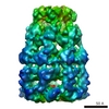
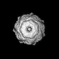
| Entry | Database: EMDB / ID: EMD-1203 | |||||||||
|---|---|---|---|---|---|---|---|---|---|---|
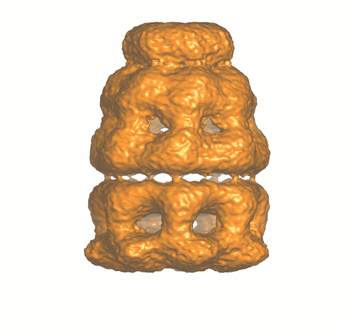
| Title | An expanded protein folding cage in the GroEL-gp31 complex. | |||||||||
 Map data Map data | GroEL-ATP-gp31 3D density map | |||||||||
 Sample Sample |
| |||||||||
| Function / homology | : / protein binding / Chaperonin Cpn60/GroEL Function and homology information Function and homology information | |||||||||
| Biological species |  | |||||||||
| Method | single particle reconstruction / cryo EM / Resolution: 12.0 Å | |||||||||
 Authors Authors | Clare DK / Bakkes PJ / van Heerikhuizen H / van der Vies SM / Saibil HR | |||||||||
 Citation Citation | Journal: J Mol Biol / Year: 2006 Title: An expanded protein folding cage in the GroEL-gp31 complex. Authors: Daniel K Clare / Patrick J Bakkes / Harm van Heerikhuizen / Saskia M van der Vies / Helen R Saibil /  Abstract: Bacteriophage T4 produces a GroES analogue, gp31, which cooperates with the Escherichia coli GroEL to fold its major coat protein gp23. We have used cryo-electron microscopy and image processing to ...Bacteriophage T4 produces a GroES analogue, gp31, which cooperates with the Escherichia coli GroEL to fold its major coat protein gp23. We have used cryo-electron microscopy and image processing to obtain three-dimensional structures of the E.coli chaperonin GroEL complexed with gp31, in the presence of both ATP and ADP. The GroEL-gp31-ADP map has a resolution of 8.2 A, which allows accurate fitting of the GroEL and gp31 crystal structures. Comparison of this fitted structure with that of the GroEL-GroES-ADP structure previously determined by cryo-electron microscopy shows that the folding cage is expanded. The enlarged volume for folding is consistent with the size of the bacteriophage coat protein gp23, which is the major substrate of GroEL-gp31 chaperonin complex. At 56 kDa, gp23 is close to the maximum size limit of a polypeptide that is thought to fit inside the GroEL-GroES folding cage. | |||||||||
| History |
|
- Structure visualization
| Movie |
Movie viewer |
|---|---|
| Structure viewer | EM map: SurfViewMolmilJmol/JSmol |
| Supplemental images |
- Downloads & links
-EMDB archive
| Map data | emd_1203.map.gz | 13.8 MB | EMDB map data format | |
|---|---|---|---|---|
| Header (meta data) | emd-1203-v30.xmlemd-1203.xml | 11.6 KB 11.6 KB | Display Display | EMDB header |
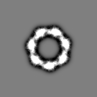
| Images |  1203.gif 1203.gif | 75.9 KB | ||
| Archive directory |  http://ftp.pdbj.org/pub/emdb/structures/EMD-1203ftp://ftp.pdbj.org/pub/emdb/structures/EMD-1203 http://ftp.pdbj.org/pub/emdb/structures/EMD-1203ftp://ftp.pdbj.org/pub/emdb/structures/EMD-1203 | HTTPS FTP |
-Validation report
| Summary document | emd_1203_validation.pdf.gz | 228.4 KB | Display | EMDB validaton report |
|---|---|---|---|---|
| Full document | emd_1203_full_validation.pdf.gz | 227.5 KB | Display | |
| Data in XML | emd_1203_validation.xml.gz | 6.1 KB | Display | |
| Arichive directory | https://ftp.pdbj.org/pub/emdb/validation_reports/EMD-1203ftp://ftp.pdbj.org/pub/emdb/validation_reports/EMD-1203 | HTTPS FTP |
-Related structure data
-Links
| EMDB pages | EMDB (EBI/PDBe) / EMDataResource |
|---|
-Map
| File | Download / File: emd_1203.map.gz / Format: CCP4 / Size: 26.4 MB / Type: IMAGE STORED AS FLOATING POINT NUMBER (4 BYTES) | ||||||||||||||||||||||||||||||||||||||||||||||||||||||||||||||||||||
|---|---|---|---|---|---|---|---|---|---|---|---|---|---|---|---|---|---|---|---|---|---|---|---|---|---|---|---|---|---|---|---|---|---|---|---|---|---|---|---|---|---|---|---|---|---|---|---|---|---|---|---|---|---|---|---|---|---|---|---|---|---|---|---|---|---|---|---|---|---|
| Annotation | GroEL-ATP-gp31 3D density map | ||||||||||||||||||||||||||||||||||||||||||||||||||||||||||||||||||||
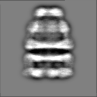
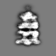
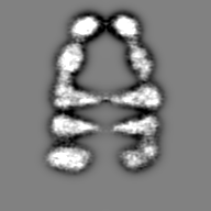
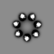
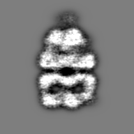
| Projections & slices | Image control
Images are generated by Spider. | ||||||||||||||||||||||||||||||||||||||||||||||||||||||||||||||||||||
| Voxel size | X=Y=Z: 1.4 Å | ||||||||||||||||||||||||||||||||||||||||||||||||||||||||||||||||||||
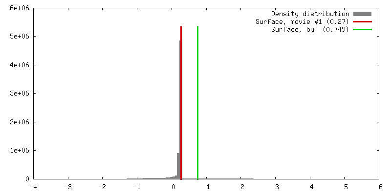
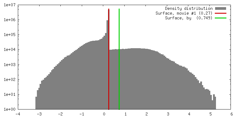
| Density |
| ||||||||||||||||||||||||||||||||||||||||||||||||||||||||||||||||||||
| Symmetry | Space group: 1 | ||||||||||||||||||||||||||||||||||||||||||||||||||||||||||||||||||||
| Details | EMDB XML:
CCP4 map header:
| ||||||||||||||||||||||||||||||||||||||||||||||||||||||||||||||||||||
 Z (Sec.)
 Y (Row.)
 X (Col.)


-Supplemental data
- Sample components
-Entire : GroEL-ATP-gp31
| Entire | Name: GroEL-ATP-gp31 |
|---|---|
| Components |
|
-Supramolecule #1000: GroEL-ATP-gp31
| Supramolecule | Name: GroEL-ATP-gp31 / type: sample / ID: 1000 Oligomeric state: heptamer of gp31 and a tetradecamer of GroEL Number unique components: 3 |
|---|---|
| Molecular weight | Experimental: 900 KDa / Theoretical: 900 KDa / Method: Mass spectrometry |
-Macromolecule #1: GroEL
| Macromolecule | Name: GroEL / type: protein_or_peptide / ID: 1 / Name.synonym: chaperonin / Number of copies: 1 / Oligomeric state: tetradecamer / Recombinant expression: Yes |
|---|---|
| Source (natural) | Organism: |
| Molecular weight | Experimental: 800 KDa / Theoretical: 800 KDa |
| Recombinant expression | Organism: |
| Sequence | GO: protein binding / InterPro: Chaperonin Cpn60/GroEL |
-Macromolecule #2: gp31
| Macromolecule | Name: gp31 / type: protein_or_peptide / ID: 2 / Name.synonym: co-chaperonin / Number of copies: 1 / Oligomeric state: heptamer / Recombinant expression: Yes |
|---|---|
| Source (natural) | Organism: |
| Molecular weight | Experimental: 100 KDa / Theoretical: 100 KDa |
| Recombinant expression | Organism: |
| Sequence | InterPro: INTERPRO: IPR011597 |
-Macromolecule #3: ATP
| Macromolecule | Name: ATP / type: ligand / ID: 3 / Name.synonym: nucleotide / Recombinant expression: No |
|---|
-Experimental details
-Structure determination
| Method | cryo EM |
|---|---|
 Processing Processing | single particle reconstruction |
| Aggregation state | particle |
-Sample preparation
| Concentration | 1 mg/mL |
|---|---|
| Buffer | pH: 7.4 / Details: 20mM Tris-HCL, 10mM MgCl, 10mM KCl |
| Grid | Details: 300 mesh copper grid - holey carbon film |
| Vitrification | Cryogen name: ETHANE / Chamber humidity: 95 % / Chamber temperature: 100 K / Instrument: HOMEMADE PLUNGER / Details: Vitrification instrument: home made / Timed resolved state: Vitrified after 5 minute incubation Method: The grids were bloted for 2-3 seconds and then left to equilibrate for 2-3 seconds and then plunged into liquid ethane |
- Electron microscopy
| Microscope | FEI TECNAI F20 |
|---|---|
| Temperature | Average: 100 K |
| Alignment procedure | Legacy - Astigmatism: objective lens astigmatism was corrected at 150kX |
| Date | Sep 1, 2004 |
| Image recording | Category: FILM / Film or detector model: KODAK SO-163 FILM / Digitization - Scanner: ZEISS SCAI / Digitization - Sampling interval: 7 µm / Number real images: 14 / Average electron dose: 15 e/Å2 / Od range: 1 / Bits/pixel: 8 |
| Electron beam | Acceleration voltage: 200 kV / Electron source:  FIELD EMISSION GUN FIELD EMISSION GUN |
| Electron optics | Calibrated magnification: 50000 / Illumination mode: FLOOD BEAM / Imaging mode: BRIGHT FIELD / Cs: 2 mm / Nominal defocus max: 3.1 µm / Nominal defocus min: 1.5 µm / Nominal magnification: 50000 |
| Sample stage | Specimen holder: single tilt / Specimen holder model: GATAN LIQUID NITROGEN |
| Experimental equipment |  Model: Tecnai F20 / Image courtesy: FEI Company |
