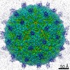
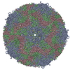
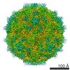
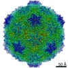
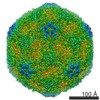
Journal: J Biol Chem / Year: 2006 Title: Structural and functional insights into the interaction of echoviruses and decay-accelerating factor. Authors: David M Pettigrew / David T Williams / David Kerrigan / David J Evans / Susan M Lea / David Bhella / Abstract: Many enteroviruses bind to the complement control protein decay-accelerating factor (DAF) to facilitate cell entry. We present here a structure for echovirus (EV) type 12 bound to DAF using cryo- ...Many enteroviruses bind to the complement control protein decay-accelerating factor (DAF) to facilitate cell entry. We present here a structure for echovirus (EV) type 12 bound to DAF using cryo-negative stain transmission electron microscopy and three-dimensional image reconstruction to 16-A resolution, which we interpreted using the atomic structures of EV11 and DAF. DAF binds to a hypervariable region of the capsid close to the 2-fold symmetry axes in an interaction that involves mostly the short consensus repeat 3 domain of DAF and the capsid protein VP2. A bulge in the density for the short consensus repeat 3 domain suggests that a loop at residues 174-180 rearranges to prevent steric collision between closely packed molecules at the 2-fold symmetry axes. Detailed analysis of receptor interactions between a variety of echoviruses and DAF using surface plasmon resonance and comparison of this structure (and our previous work; Bhella, D., Goodfellow, I. G., Roversi, P., Pettigrew, D., Chaudhry, Y., Evans, D. J., and Lea, S. M. (2004) J. Biol. Chem. 279, 8325-8332) with reconstructions published for EV7 bound to DAF support major differences in receptor recognition among these viruses. However, comparison of the electron density for the two virus.receptor complexes (rather than comparisons of the pseudo-atomic models derived from fitting the coordinates into these densities) suggests that the dramatic differences in interaction affinities/specificities may arise from relatively subtle structural differences rather than from large-scale repositioning of the receptor with respect to the virus surface.
History
Deposition
Dec 5, 2005
-
Header (metadata) release
Dec 5, 2005
-
Map release
Dec 5, 2005
-
Update
May 26, 2011
-
Current status
May 26, 2011
Processing site: PDBe / Status: Released
-
Structure visualization
Movie
Surface view with section colored by density value
Name: Echovirus type 12 / type: sample / ID: 1000 / Number unique components: 1
-
Supramolecule #1: Human echovirus 12
Supramolecule
Name: Human echovirus 12 / type: virus / ID: 1 / Name.synonym: EV12 / NCBI-ID: 35293 / Sci species name: Human echovirus 12 / Virus type: VIRION / Virus isolate: SEROTYPE / Virus enveloped: No / Virus empty: No / Syn species name: EV12
Host (natural)
Organism: Homo sapiens (human) / synonym: VERTEBRATES
Virus shell
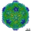
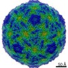
Shell ID: 1 / Name: VP1-4 / Diameter: 300 Å / T number (triangulation number): 3
-
Experimental details
-
Structure determination
Method
negative staining, cryo EM
Processing
single particle reconstruction
Aggregation state
particle
-
Sample preparation
Concentration
0.2 mg/mL
Buffer
pH: 7.4 / Details: Phosphate buffered saline
Staining
Type: NEGATIVE Details: 5 microlitres of sample was loaded onto a quantifoil grid and then floated on a 20 microlitre droplet of 15% Ammonium Molybdate for 10 seconds, then the grid was blotted for 2 seconds before ...Details: 5 microlitres of sample was loaded onto a quantifoil grid and then floated on a 20 microlitre droplet of 15% Ammonium Molybdate for 10 seconds, then the grid was blotted for 2 seconds before vitrification in liquid ethane.
Grid
Details: 400 mesh R2/2 quantifoils
Vitrification
Cryogen name: ETHANE / Method: Blot for two seconds, wait for two seconds.
-
Electron microscopy
Microscope
JEOL 1200
Temperature
Average: 100 K
Alignment procedure
Legacy - Astigmatism: Objective astigmatism corrected at 200,000 times mag
Image recording
Category: FILM / Film or detector model: KODAK SO-163 FILM / Digitization - Scanner: NIKON COOLSCAN / Digitization - Sampling interval: 6.35 µm / Number real images: 84 / Details: Scanned on Nikon Coolscan / Bits/pixel: 16
Tilt angle min
0
Tilt angle max
0
Electron beam
Acceleration voltage: 120 kV / Electron source: LAB6
In the structure databanks used in Yorodumi, some data are registered as the other names, "COVID-19 virus" and "2019-nCoV". Here are the details of the virus and the list of structure data.
Jan 31, 2019. EMDB accession codes are about to change! (news from PDBe EMDB page)
EMDB accession codes are about to change! (news from PDBe EMDB page)
The allocation of 4 digits for EMDB accession codes will soon come to an end. Whilst these codes will remain in use, new EMDB accession codes will include an additional digit and will expand incrementally as the available range of codes is exhausted. The current 4-digit format prefixed with “EMD-” (i.e. EMD-XXXX) will advance to a 5-digit format (i.e. EMD-XXXXX), and so on. It is currently estimated that the 4-digit codes will be depleted around Spring 2019, at which point the 5-digit format will come into force.
The EM Navigator/Yorodumi systems omit the EMD- prefix.
Related info.:Q: What is EMD? / ID/Accession-code notation in Yorodumi/EM Navigator
Yorodumi is a browser for structure data from EMDB, PDB, SASBDB, etc.
This page is also the successor to EM Navigator detail page, and also detail information page/front-end page for Omokage search.
The word "yorodu" (or yorozu) is an old Japanese word meaning "ten thousand". "mi" (miru) is to see.
Related info.:EMDB / PDB / SASBDB / Comparison of 3 databanks / Yorodumi Search / Aug 31, 2016. New EM Navigator & Yorodumi / Yorodumi Papers / Jmol/JSmol / Function and homology information / Changes in new EM Navigator and Yorodumi
 Movie
Movie Controller

 Yorodumi
 Open data
 Basic information
 Map data
 Sample
 Function and homology information
 Human echovirus 12
 Authors
 Citation
 Structure visualization
 UCSF Chimera
 Downloads & links
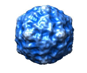 1183.gif
 http://ftp.pdbj.org/pub/emdb/structures/EMD-1183


 Z (Sec.)
 Y (Row.)
 X (Col.)


 Sample components
 Homo sapiens (human) / synonym: VERTEBRATES
 Processing
 Electron microscopy