[English] 日本語
 Yorodumi
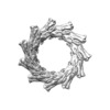
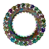
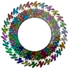
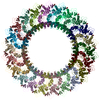
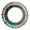
- EMDB-11468: C11 symmetry: Bacterial Vipp1 and PspA are members of the ancient... -
+ Open data
- Basic information
| Entry | Database: EMDB / ID: EMD-11468 | |||||||||
|---|---|---|---|---|---|---|---|---|---|---|
| Title | C11 symmetry: Bacterial Vipp1 and PspA are members of the ancient ESCRT-III membrane-remodeling superfamily. | |||||||||
 Map data Map data | ||||||||||
 Sample Sample |
| |||||||||
 Keywords Keywords | membrane remodelling / LIPID BINDING PROTEIN | |||||||||
| Function / homology | PspA/IM30 / PspA/IM30 family / lipid binding / plasma membrane / Membrane-associated protein Vipp1 Function and homology information Function and homology information | |||||||||
| Biological species |  Nostoc punctiforme (bacteria) Nostoc punctiforme (bacteria) | |||||||||
| Method | single particle reconstruction / cryo EM / Resolution: 8.2 Å | |||||||||
 Authors Authors | Liu J / Tassinari M / Souza DP / Naskar S / Noel JK / Bohuszewicz O / Buck M / Williams TA / Baum B / Low HH | |||||||||
| Funding support |  United Kingdom, 1 items United Kingdom, 1 items
| |||||||||
 Citation Citation |  Journal: Cell / Year: 2021 Journal: Cell / Year: 2021Title: Bacterial Vipp1 and PspA are members of the ancient ESCRT-III membrane-remodeling superfamily. Authors: Jiwei Liu / Matteo Tassinari / Diorge P Souza / Souvik Naskar / Jeffrey K Noel / Olga Bohuszewicz / Martin Buck / Tom A Williams / Buzz Baum / Harry H Low /   Abstract: Membrane remodeling and repair are essential for all cells. Proteins that perform these functions include Vipp1/IM30 in photosynthetic plastids, PspA in bacteria, and ESCRT-III in eukaryotes. Here, ...Membrane remodeling and repair are essential for all cells. Proteins that perform these functions include Vipp1/IM30 in photosynthetic plastids, PspA in bacteria, and ESCRT-III in eukaryotes. Here, using a combination of evolutionary and structural analyses, we show that these protein families are homologous and share a common ancient evolutionary origin that likely predates the last universal common ancestor. This homology is evident in cryo-electron microscopy structures of Vipp1 rings from the cyanobacterium Nostoc punctiforme presented over a range of symmetries. Each ring is assembled from rungs that stack and progressively tilt to form dome-shaped curvature. Assembly is facilitated by hinges in the Vipp1 monomer, similar to those in ESCRT-III proteins, which allow the formation of flexible polymers. Rings have an inner lumen that is able to bind and deform membranes. Collectively, these data suggest conserved mechanistic principles that underlie Vipp1, PspA, and ESCRT-III-dependent membrane remodeling across all domains of life. | |||||||||
| History |
|
- Structure visualization
| Movie |
 Movie viewer Movie viewer |
|---|---|
| Structure viewer | EM map:  SurfView SurfView Molmil Molmil Jmol/JSmol Jmol/JSmol |
| Supplemental images |
- Downloads & links
-EMDB archive
| Map data |  emd_11468.map.gz emd_11468.map.gz | 11.8 MB |  EMDB map data format EMDB map data format | |
|---|---|---|---|---|
| Header (meta data) |  emd-11468-v30.xml emd-11468-v30.xml emd-11468.xml emd-11468.xml | 9.2 KB 9.2 KB | Display Display |  EMDB header EMDB header |
| Images |  emd_11468.png emd_11468.png | 35.3 KB | ||
| Filedesc metadata |  emd-11468.cif.gz emd-11468.cif.gz | 4.8 KB | ||
| Archive directory |  http://ftp.pdbj.org/pub/emdb/structures/EMD-11468 http://ftp.pdbj.org/pub/emdb/structures/EMD-11468 ftp://ftp.pdbj.org/pub/emdb/structures/EMD-11468 ftp://ftp.pdbj.org/pub/emdb/structures/EMD-11468 | HTTPS FTP |
-Validation report
| Summary document |  emd_11468_validation.pdf.gz emd_11468_validation.pdf.gz | 372.3 KB | Display |  EMDB validaton report EMDB validaton report |
|---|---|---|---|---|
| Full document |  emd_11468_full_validation.pdf.gz emd_11468_full_validation.pdf.gz | 371.9 KB | Display | |
| Data in XML |  emd_11468_validation.xml.gz emd_11468_validation.xml.gz | 6.7 KB | Display | |
| Data in CIF |  emd_11468_validation.cif.gz emd_11468_validation.cif.gz | 7.7 KB | Display | |
| Arichive directory |  https://ftp.pdbj.org/pub/emdb/validation_reports/EMD-11468 https://ftp.pdbj.org/pub/emdb/validation_reports/EMD-11468 ftp://ftp.pdbj.org/pub/emdb/validation_reports/EMD-11468 ftp://ftp.pdbj.org/pub/emdb/validation_reports/EMD-11468 | HTTPS FTP |
-Related structure data
| Related structure data |  6zvrMC  6zvsC  6zvtC  6zw4C  6zw5C  6zw6C  6zw7C M: atomic model generated by this map C: citing same article ( |
|---|---|
| Similar structure data |
- Links
Links
| EMDB pages |  EMDB (EBI/PDBe) / EMDB (EBI/PDBe) /  EMDataResource EMDataResource |
|---|
- Map
Map
| File |  Download / File: emd_11468.map.gz / Format: CCP4 / Size: 144.7 MB / Type: IMAGE STORED AS FLOATING POINT NUMBER (4 BYTES) Download / File: emd_11468.map.gz / Format: CCP4 / Size: 144.7 MB / Type: IMAGE STORED AS FLOATING POINT NUMBER (4 BYTES) | ||||||||||||||||||||||||||||||||||||||||||||||||||||||||||||
|---|---|---|---|---|---|---|---|---|---|---|---|---|---|---|---|---|---|---|---|---|---|---|---|---|---|---|---|---|---|---|---|---|---|---|---|---|---|---|---|---|---|---|---|---|---|---|---|---|---|---|---|---|---|---|---|---|---|---|---|---|---|
| Projections & slices | Image control
Images are generated by Spider. | ||||||||||||||||||||||||||||||||||||||||||||||||||||||||||||
| Voxel size | X=Y=Z: 1.39 Å | ||||||||||||||||||||||||||||||||||||||||||||||||||||||||||||
| Density |
| ||||||||||||||||||||||||||||||||||||||||||||||||||||||||||||
| Symmetry | Space group: 1 | ||||||||||||||||||||||||||||||||||||||||||||||||||||||||||||
| Details | EMDB XML:
CCP4 map header:
| ||||||||||||||||||||||||||||||||||||||||||||||||||||||||||||
-Supplemental data
- Sample components
-Entire : vipp1 c11 ring
| Entire | Name: vipp1 c11 ring |
|---|---|
| Components |
|
-Supramolecule #1: vipp1 c11 ring
| Supramolecule | Name: vipp1 c11 ring / type: complex / ID: 1 / Parent: 0 / Macromolecule list: all |
|---|---|
| Source (natural) | Organism:  Nostoc punctiforme (bacteria) Nostoc punctiforme (bacteria) |
-Macromolecule #1: Vipp1
| Macromolecule | Name: Vipp1 / type: protein_or_peptide / ID: 1 / Number of copies: 55 / Enantiomer: LEVO |
|---|---|
| Source (natural) | Organism:  Nostoc punctiforme (bacteria) Nostoc punctiforme (bacteria) |
| Molecular weight | Theoretical: 28.745461 KDa |
| Recombinant expression | Organism:  |
| Sequence | String: MGLFDRIKRV VSSNLNDLVN KAEDPEKMLE QAILEMQEDL VQLRQGVAQA IAAQKRSEKQ YNDAQNEINK WQRNAQLALQ KGDENLARQ ALERKKTYTD TSAALKASLD TQSTQVETLK RNLIQLESKI SEAKTKKEML KARITTAKAQ EQLQGMVRGM N TSSAMSAF ...String: MGLFDRIKRV VSSNLNDLVN KAEDPEKMLE QAILEMQEDL VQLRQGVAQA IAAQKRSEKQ YNDAQNEINK WQRNAQLALQ KGDENLARQ ALERKKTYTD TSAALKASLD TQSTQVETLK RNLIQLESKI SEAKTKKEML KARITTAKAQ EQLQGMVRGM N TSSAMSAF ERMEEKVLMQ ESRAQALGEL AGADLETQFA QLEGGSDVDD ELAALKAQML PPATPVTQAQ LPPQQETTPA KS NEVVDAE LDSLRKQLDQ L UniProtKB: Membrane-associated protein Vipp1 |
-Experimental details
-Structure determination
| Method | cryo EM |
|---|---|
 Processing Processing | single particle reconstruction |
| Aggregation state | particle |
- Sample preparation
| Buffer | pH: 8.4 |
|---|---|
| Vitrification | Cryogen name: ETHANE |
- Electron microscopy
| Microscope | FEI TITAN KRIOS |
|---|---|
| Image recording | Film or detector model: GATAN K2 SUMMIT (4k x 4k) / Average electron dose: 1.5 e/Å2 |
| Electron beam | Acceleration voltage: 300 kV / Electron source:  FIELD EMISSION GUN FIELD EMISSION GUN |
| Electron optics | Illumination mode: FLOOD BEAM / Imaging mode: BRIGHT FIELD |
| Experimental equipment |  Model: Titan Krios / Image courtesy: FEI Company |
- Image processing
| Startup model | Type of model: NONE |
|---|---|
| Final reconstruction | Resolution.type: BY AUTHOR / Resolution: 8.2 Å / Resolution method: FSC 0.143 CUT-OFF / Software - Name: RELION / Number images used: 3912 |
| Initial angle assignment | Type: NOT APPLICABLE |
| Final angle assignment | Type: NOT APPLICABLE |
 Movie
Movie Controller






 Z (Sec.)
 Y (Row.)
 X (Col.)


