 Movie
Movie Controller

[English] 日本語
 Yorodumi
- EMDB-11413: Structure of left-handed protein cage consisting of 24 eleven-mem... -
+ Open data

- Basic information
| Entry | Database: EMDB / ID: EMD-11413 | |||||||||
|---|---|---|---|---|---|---|---|---|---|---|
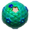
| Title | Structure of left-handed protein cage consisting of 24 eleven-membered ring proteins held together by BMH cross linkers | |||||||||
 Map data Map data | Structure of left-handed protein cage consisting of 24 eleven-membered ring proteins held together by BMH cross linkers | |||||||||
 Sample Sample |
| |||||||||
| Biological species |   Geobacillus stearothermophilus (bacteria) Geobacillus stearothermophilus (bacteria) | |||||||||
| Method | single particle reconstruction / cryo EM / Resolution: 6.8 Å | |||||||||
 Authors Authors | Biela AP / Maskell D | |||||||||
| Funding support |  Poland, 1 items Poland, 1 items
| |||||||||
 Citation Citation | Journal: Sci Adv / Year: 2022 Title: Chemically induced protein cage assembly with programmable opening and cargo release. Authors: Izabela Stupka / Yusuke Azuma / Artur P Biela / Motonori Imamura / Simon Scheuring / Elżbieta Pyza / Olga Woźnicka / Daniel P Maskell / Jonathan G Heddle /   Abstract: Engineered protein cages are promising tools that can be customized for applications in medicine and nanotechnology. A major challenge is developing a straightforward strategy for endowing cages with ...Engineered protein cages are promising tools that can be customized for applications in medicine and nanotechnology. A major challenge is developing a straightforward strategy for endowing cages with bespoke, inducible disassembly. Such cages would allow release of encapsulated cargoes at desired timing and location. Here, we achieve such programmable disassembly using protein cages, in which the subunits are held together by different molecular cross-linkers. This modular system enables cage disassembly to be controlled in a condition-dependent manner. Structural details of the resulting cages were determined using cryo–electron microscopy, which allowed observation of bridging cross-linkers at intended positions. Triggered disassembly was demonstrated by high-speed atomic force microscopy and subsequent cargo release using an encapsulated Förster resonance energy transfer pair whose signal depends on the quaternary structure of the cage. | |||||||||
| History |
|
- Structure visualization
| Movie |
 Movie viewer Movie viewer |
|---|---|
| Structure viewer | EM map: SurfViewMolmilJmol/JSmol |
| Supplemental images |

- Downloads & links
-EMDB archive
| Map data | emd_11413.map.gz | 38.1 MB | EMDB map data format | |
|---|---|---|---|---|
| Header (meta data) | emd-11413-v30.xmlemd-11413.xml | 17.1 KB 17.1 KB | Display Display | EMDB header |
| FSC (resolution estimation) | emd_11413_fsc.xml | 7.9 KB | Display | FSC data file |
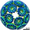
| Images | 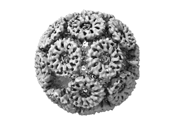 emd_11413.png emd_11413.png | 89.2 KB | ||
| Masks | emd_11413_msk_1.map | 40.6 MB | Mask map | |
| Others | emd_11413_half_map_1.map.gzemd_11413_half_map_2.map.gz | 37.2 MB 37.2 MB | ||
| Archive directory |  http://ftp.pdbj.org/pub/emdb/structures/EMD-11413ftp://ftp.pdbj.org/pub/emdb/structures/EMD-11413 http://ftp.pdbj.org/pub/emdb/structures/EMD-11413ftp://ftp.pdbj.org/pub/emdb/structures/EMD-11413 | HTTPS FTP |
-Related structure data
| Related structure data | C: citing same article ( |
|---|---|
| Similar structure data |


-Links
| EMDB pages | EMDB (EBI/PDBe) / EMDataResource |
|---|---|
| Related items in Molecule of the Month |

-Map
| File | Download / File: emd_11413.map.gz / Format: CCP4 / Size: 40.6 MB / Type: IMAGE STORED AS FLOATING POINT NUMBER (4 BYTES) | ||||||||||||||||||||||||||||||||||||||||||||||||||||||||||||
|---|---|---|---|---|---|---|---|---|---|---|---|---|---|---|---|---|---|---|---|---|---|---|---|---|---|---|---|---|---|---|---|---|---|---|---|---|---|---|---|---|---|---|---|---|---|---|---|---|---|---|---|---|---|---|---|---|---|---|---|---|---|
| Annotation | Structure of left-handed protein cage consisting of 24 eleven-membered ring proteins held together by BMH cross linkers | ||||||||||||||||||||||||||||||||||||||||||||||||||||||||||||
| Projections & slices | Image control
Images are generated by Spider. | ||||||||||||||||||||||||||||||||||||||||||||||||||||||||||||
| Voxel size | X=Y=Z: 1.626 Å | ||||||||||||||||||||||||||||||||||||||||||||||||||||||||||||
| Density |
| ||||||||||||||||||||||||||||||||||||||||||||||||||||||||||||
| Symmetry | Space group: 1 | ||||||||||||||||||||||||||||||||||||||||||||||||||||||||||||
| Details | EMDB XML:
CCP4 map header:
| ||||||||||||||||||||||||||||||||||||||||||||||||||||||||||||
 Z (Sec.)
 Y (Row.)
 X (Col.)


-Supplemental data
-Mask #1
| File | emd_11413_msk_1.map | ||||||||||||
|---|---|---|---|---|---|---|---|---|---|---|---|---|---|
| Projections & Slices |
| ||||||||||||
| Density Histograms |


-Half map: half map A
| File | emd_11413_half_map_1.map | ||||||||||||
|---|---|---|---|---|---|---|---|---|---|---|---|---|---|
| Annotation | half map A | ||||||||||||
| Projections & Slices |
| ||||||||||||
| Density Histograms |


-Half map: half map B
| File | emd_11413_half_map_2.map | ||||||||||||
|---|---|---|---|---|---|---|---|---|---|---|---|---|---|
| Annotation | half map B | ||||||||||||
| Projections & Slices |
| ||||||||||||
| Density Histograms |


- Sample components
-Entire : chemically induced artificial protein cage
| Entire | Name: chemically induced artificial protein cage |
|---|---|
| Components |
|
-Supramolecule #1: chemically induced artificial protein cage
| Supramolecule | Name: chemically induced artificial protein cage / type: complex / ID: 1 / Parent: 0 / Macromolecule list: all / Details: BMH cross linked protein cage |
|---|---|
| Source (natural) | Organism: Geobacillus stearothermophilus (bacteria) |
| Recombinant expression | Organism: |
| Molecular weight | Experimental: 2.2 MDa |
-Macromolecule #1: Transcription attenuation protein MtrB
| Macromolecule | Name: Transcription attenuation protein MtrB / type: protein_or_peptide / ID: 1 / Enantiomer: LEVO |
|---|---|
| Source (natural) | Organism: Geobacillus stearothermophilus (bacteria) |
| Recombinant expression | Organism: |
| Sequence | String: MYTNSDFVVI KALEDGVNVI GLTRGADTRF HHSECLDKGE VLIAQFTEHT SAIKVRGKAY IQTSHGVIES EGKK |
-Experimental details
-Structure determination
| Method | cryo EM |
|---|---|
 Processing Processing | single particle reconstruction |
| Aggregation state | particle |
-Sample preparation
| Concentration | 8.9 mg/mL |
|---|---|
| Buffer | pH: 7.2 |
| Grid | Model: Quantifoil R1.2/1.3 / Material: COPPER / Mesh: 300 / Support film - Material: CARBON / Support film - topology: HOLEY / Pretreatment - Type: GLOW DISCHARGE / Pretreatment - Atmosphere: AIR |
| Vitrification | Cryogen name: ETHANE / Chamber humidity: 100 % / Chamber temperature: 277 K / Instrument: FEI VITROBOT MARK IV |
- Electron microscopy
| Microscope | FEI TITAN KRIOS |
|---|---|
| Image recording | Film or detector model: FEI FALCON III (4k x 4k) / Detector mode: COUNTING / Number grids imaged: 1 / Number real images: 10169 / Average exposure time: 1.0 sec. / Average electron dose: 35.0 e/Å2 |
| Electron beam | Acceleration voltage: 300 kV / Electron source:  FIELD EMISSION GUN FIELD EMISSION GUN |
| Electron optics | Illumination mode: FLOOD BEAM / Imaging mode: BRIGHT FIELD / Cs: 2.7 mm / Nominal magnification: 75000 |
| Sample stage | Specimen holder model: FEI TITAN KRIOS AUTOGRID HOLDER / Cooling holder cryogen: NITROGEN |
| Experimental equipment |  Model: Titan Krios / Image courtesy: FEI Company |

