 Movie
Movie Controller

[English] 日本語
 Yorodumi
- EMDB-11311: Structure of the Salmonella PrgI needle filament attached to the ... -
+ Open data

- Basic information
| Entry | Database: EMDB / ID: EMD-11311 | |||||||||
|---|---|---|---|---|---|---|---|---|---|---|
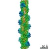
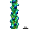
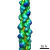
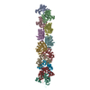
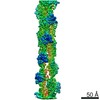
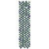
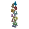
| Title | Structure of the Salmonella PrgI needle filament attached to the basal body | |||||||||
 Map data Map data | ||||||||||
 Sample Sample |
| |||||||||
 Keywords Keywords | Type 3 secretion system / Salmonella / Helical reconstruction / Needle filament. / PROTEIN TRANSPORT | |||||||||
| Function / homology |  Function and homology information Function and homology informationtype III protein secretion system complex / protein secretion by the type III secretion system / cell surface / extracellular region / identical protein binding Similarity search - Function | |||||||||
| Biological species |  Salmonella enterica subsp. enterica serovar Typhimurium (bacteria) / Salmonella typhimurium (bacteria) Salmonella enterica subsp. enterica serovar Typhimurium (bacteria) / Salmonella typhimurium (bacteria) | |||||||||
| Method | helical reconstruction / cryo EM / Resolution: 3.6 Å | |||||||||
 Authors Authors | Kotov V / Lunelli M / Wald J / Kolbe M / Marlovits TC | |||||||||
 Citation Citation | Journal: Biochem Biophys Rep / Year: 2021 Title: Helical reconstruction of and needle filaments attached to type 3 basal bodies. Authors: Vadim Kotov / Michele Lunelli / Jiri Wald / Michael Kolbe / Thomas C Marlovits /  Abstract: Gram-negative pathogens evolved a syringe-like nanomachine, termed type 3 secretion system, to deliver protein effectors into the cytoplasm of host cells. An essential component of this system is a ...Gram-negative pathogens evolved a syringe-like nanomachine, termed type 3 secretion system, to deliver protein effectors into the cytoplasm of host cells. An essential component of this system is a long helical needle filament that protrudes from the bacterial surface and connects the cytoplasms of the bacterium and the eukaryotic cell. Previous structural research was predominantly focused on reconstituted type 3 needle filaments, which lacked the biological context. In this work we introduce a facile procedure to obtain high-resolution cryo-EM structure of needle filaments attached to the basal body of type 3 secretion systems. We validate our approach by solving the structure of PrgI filament and demonstrate its utility by obtaining the first high-resolution cryo-EM reconstruction of Shigella MxiH filament. Our work paves the way to systematic structural characterization of attached type 3 needle filaments in the context of mutagenesis studies, protein structural evolution and drug development. | |||||||||
| History |
|
- Structure visualization
| Movie |
Movie viewer |
|---|---|
| Structure viewer | EM map: SurfViewMolmilJmol/JSmol |
| Supplemental images |

- Downloads & links
-EMDB archive
| Map data | emd_11311.map.gz | 96.3 MB | EMDB map data format | |
|---|---|---|---|---|
| Header (meta data) | emd-11311-v30.xmlemd-11311.xml | 16 KB 16 KB | Display Display | EMDB header |
| FSC (resolution estimation) | emd_11311_fsc.xml | 10.7 KB | Display | FSC data file |
| Images |  emd_11311.png emd_11311.png | 147.3 KB | ||
| Filedesc metadata | emd-11311.cif.gz | 5.5 KB | ||
| Others | emd_11311_half_map_1.map.gzemd_11311_half_map_2.map.gz | 80.8 MB 80.8 MB | ||
| Archive directory |  http://ftp.pdbj.org/pub/emdb/structures/EMD-11311ftp://ftp.pdbj.org/pub/emdb/structures/EMD-11311 http://ftp.pdbj.org/pub/emdb/structures/EMD-11311ftp://ftp.pdbj.org/pub/emdb/structures/EMD-11311 | HTTPS FTP |
-Related structure data
| Related structure data |  6znhMC  6zniC M: atomic model generated by this map C: citing same article ( |
|---|---|
| Similar structure data |


-Links
| EMDB pages | EMDB (EBI/PDBe) / EMDataResource |
|---|
-Map
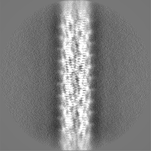
| File | Download / File: emd_11311.map.gz / Format: CCP4 / Size: 103 MB / Type: IMAGE STORED AS FLOATING POINT NUMBER (4 BYTES) | ||||||||||||||||||||||||||||||||||||||||||||||||||||||||||||
|---|---|---|---|---|---|---|---|---|---|---|---|---|---|---|---|---|---|---|---|---|---|---|---|---|---|---|---|---|---|---|---|---|---|---|---|---|---|---|---|---|---|---|---|---|---|---|---|---|---|---|---|---|---|---|---|---|---|---|---|---|---|
| Projections & slices | Image control
Images are generated by Spider. | ||||||||||||||||||||||||||||||||||||||||||||||||||||||||||||
| Voxel size | X=Y=Z: 1.09 Å | ||||||||||||||||||||||||||||||||||||||||||||||||||||||||||||
| Density |
| ||||||||||||||||||||||||||||||||||||||||||||||||||||||||||||
| Symmetry | Space group: 1 | ||||||||||||||||||||||||||||||||||||||||||||||||||||||||||||
| Details | EMDB XML:
CCP4 map header:
| ||||||||||||||||||||||||||||||||||||||||||||||||||||||||||||
 Z (Sec.)
 Y (Row.)
 X (Col.)


-Supplemental data
-Half map: #1
| File | emd_11311_half_map_1.map | ||||||||||||
|---|---|---|---|---|---|---|---|---|---|---|---|---|---|
| Projections & Slices |
| ||||||||||||
| Density Histograms |


-Half map: #2
| File | emd_11311_half_map_2.map | ||||||||||||
|---|---|---|---|---|---|---|---|---|---|---|---|---|---|
| Projections & Slices |
| ||||||||||||
| Density Histograms |


- Sample components
-Entire : Needle filament of the type III secretion system
| Entire | Name: Needle filament of the type III secretion system |
|---|---|
| Components |
|
-Supramolecule #1: Needle filament of the type III secretion system
| Supramolecule | Name: Needle filament of the type III secretion system / type: organelle_or_cellular_component / ID: 1 / Parent: 0 / Macromolecule list: all Details: Structure obtained from needles attached to the basal body |
|---|---|
| Source (natural) | Organism: Salmonella enterica subsp. enterica serovar Typhimurium (bacteria) Strain: SB905 |
-Macromolecule #1: PrgI
| Macromolecule | Name: PrgI / type: protein_or_peptide / ID: 1 / Number of copies: 23 / Enantiomer: LEVO |
|---|---|
| Source (natural) | Organism: Salmonella typhimurium (bacteria) |
| Molecular weight | Theoretical: 8.864868 KDa |
| Sequence | String: MATPWSGYLD DVSAKFDTGV DNLQTQVTEA LDKLAAKPSD PALLAAYQSK LSEYNLYRNA QSNTVKVFKD IDAAIIQNFR UniProtKB: SPI-1 type 3 secretion system needle filament protein |
-Experimental details
-Structure determination
| Method | cryo EM |
|---|---|
 Processing Processing | helical reconstruction |
| Aggregation state | particle |
-Sample preparation
| Buffer | pH: 8 Component:
| |||||||||||||||
|---|---|---|---|---|---|---|---|---|---|---|---|---|---|---|---|---|
| Grid | Model: Quantifoil / Material: COPPER / Support film - Material: GRAPHENE OXIDE / Support film - topology: CONTINUOUS / Pretreatment - Type: GLOW DISCHARGE | |||||||||||||||
| Vitrification | Cryogen name: ETHANE-PROPANE / Chamber humidity: 100 % / Instrument: FEI VITROBOT MARK IV |
- Electron microscopy
| Microscope | FEI TITAN KRIOS |
|---|---|
| Specialist optics | Energy filter - Name: GIF Bioquantum |
| Image recording | Film or detector model: GATAN K2 SUMMIT (4k x 4k) / Detector mode: COUNTING / Average exposure time: 5.0 sec. / Average electron dose: 31.0 e/Å2 |
| Electron beam | Acceleration voltage: 300 kV / Electron source:  FIELD EMISSION GUN FIELD EMISSION GUN |
| Electron optics | Illumination mode: FLOOD BEAM / Imaging mode: BRIGHT FIELD / Cs: 2.7 mm / Nominal defocus max: 3.5 µm / Nominal defocus min: 0.8 µm / Nominal magnification: 130000 |
| Sample stage | Specimen holder model: FEI TITAN KRIOS AUTOGRID HOLDER / Cooling holder cryogen: NITROGEN |
| Experimental equipment |  Model: Titan Krios / Image courtesy: FEI Company |


