 ムービー
ムービー コントローラー


+ データを開く

- 基本情報
基本情報
| 登録情報 | データベース: EMDB / ID: EMD-11304 | |||||||||
|---|---|---|---|---|---|---|---|---|---|---|
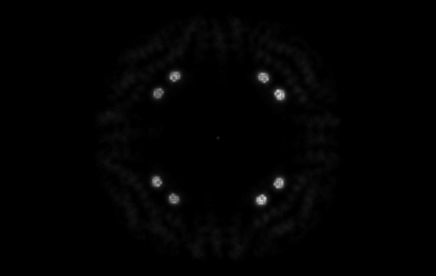
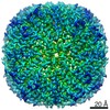
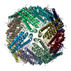
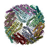
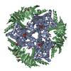
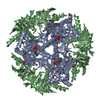
| タイトル | radiation damage-assisted analysis of iron-filled ferritin-M6A. | |||||||||
 マップデータ マップデータ | radiation damage-assisted analysis of Iron sediments in Ferritin-M6A | |||||||||
 試料 試料 |
| |||||||||
| 機能・相同性 |  機能・相同性情報 機能・相同性情報intracellular sequestering of iron ion / ferric iron binding / ferrous iron binding / iron ion transport / cytoplasm 類似検索 - 分子機能 | |||||||||
| 生物種 |  | |||||||||
| 手法 | 単粒子再構成法 / クライオ電子顕微鏡法 / 解像度: 3.5 Å | |||||||||
 データ登録者 データ登録者 | Davidov G / Abelya G / Zalk R / Izbicki B / Shaibi S / Spektor L / Shagidov D / Meyron Holtz EG / Zarivach R / Frank GA | |||||||||
| 資金援助 |  イスラエル, 2件 イスラエル, 2件
| |||||||||
 引用 引用 | ジャーナル: J Am Chem Soc / 年: 2020 タイトル: Folding of an Intrinsically Disordered Iron-Binding Peptide in Response to Sedimentation Revealed by Cryo-EM. 著者: Geula Davidov / Gili Abelya / Ran Zalk / Benjamin Izbicki / Sharon Shaibi / Lior Spektor / Dayana Shagidov / Esther G Meyron-Holtz / Raz Zarivach / Gabriel A Frank / 要旨: Biomineralization is mediated by specialized proteins that guide and control mineral sedimentation. In many cases, the active regions of these biomineralization proteins are intrinsically disordered. ...Biomineralization is mediated by specialized proteins that guide and control mineral sedimentation. In many cases, the active regions of these biomineralization proteins are intrinsically disordered. High-resolution structures of these proteins while they interact with minerals are essential for understanding biomineralization processes and the function of intrinsically disordered proteins (IDPs). Here we used the cavity of ferritin as a nanoreactor where the interaction between M6A, an intrinsically disordered iron-binding domain, and an iron oxide particle was visualized at high resolution by cryo-EM. Taking advantage of the differences in the electron-dose sensitivity of the protein and the iron oxide particles, we developed a method to determine the irregular shape of the particles found in our density maps. We found that the folding of M6A correlates with the detection of mineral particles in its vicinity. M6A interacts with the iron oxide particles through its C-terminal side, resulting in the stabilization of a helix at its N-terminal side. The stabilization of the helix at a region that is not in direct contact with the iron oxide particle demonstrates the ability of IDPs to respond to signals from their surroundings by conformational changes. These findings provide the first glimpse toward the long-suspected mechanism for biomineralization protein control over mineral microstructure, where unstructured regions of these proteins become more ordered in response to their interaction with the nascent mineral particles. | |||||||||
| 履歴 |
|
- 構造の表示
構造の表示
| ムービー |
ムービービューア |
|---|---|
| 構造ビューア | EMマップ: SurfViewMolmilJmol/JSmol |
| 添付画像 |
- ダウンロードとリンク
-EMDBアーカイブ
| マップデータ | emd_11304.map.gz | 45.5 MB | EMDBマップデータ形式 | |
|---|---|---|---|---|
| ヘッダ (付随情報) | emd-11304-v30.xmlemd-11304.xml | 11 KB 11 KB | 表示 表示 | EMDBヘッダ |
| 画像 |  emd_11304.png emd_11304.png | 33.5 KB | ||
| アーカイブディレクトリ |  http://ftp.pdbj.org/pub/emdb/structures/EMD-11304ftp://ftp.pdbj.org/pub/emdb/structures/EMD-11304 http://ftp.pdbj.org/pub/emdb/structures/EMD-11304ftp://ftp.pdbj.org/pub/emdb/structures/EMD-11304 | HTTPS FTP |
-検証レポート
| 文書・要旨 | emd_11304_validation.pdf.gz | 295 KB | 表示 | EMDB検証レポート |
|---|---|---|---|---|
| 文書・詳細版 | emd_11304_full_validation.pdf.gz | 294.6 KB | 表示 | |
| XML形式データ | emd_11304_validation.xml.gz | 5.9 KB | 表示 | |
| CIF形式データ | emd_11304_validation.cif.gz | 6.7 KB | 表示 | |
| アーカイブディレクトリ | https://ftp.pdbj.org/pub/emdb/validation_reports/EMD-11304ftp://ftp.pdbj.org/pub/emdb/validation_reports/EMD-11304 | HTTPS FTP |
-関連構造データ


-リンク
| EMDBのページ | EMDB (EBI/PDBe) / EMDataResource |
|---|---|
| 「今月の分子」の関連する項目 |


-マップ
| ファイル | ダウンロード / ファイル: emd_11304.map.gz / 形式: CCP4 / 大きさ: 64 MB / タイプ: IMAGE STORED AS FLOATING POINT NUMBER (4 BYTES) | ||||||||||||||||||||||||||||||||||||||||||||||||||||||||||||
|---|---|---|---|---|---|---|---|---|---|---|---|---|---|---|---|---|---|---|---|---|---|---|---|---|---|---|---|---|---|---|---|---|---|---|---|---|---|---|---|---|---|---|---|---|---|---|---|---|---|---|---|---|---|---|---|---|---|---|---|---|---|
| 注釈 | radiation damage-assisted analysis of Iron sediments in Ferritin-M6A | ||||||||||||||||||||||||||||||||||||||||||||||||||||||||||||
| ボクセルのサイズ | X=Y=Z: 1.1 Å | ||||||||||||||||||||||||||||||||||||||||||||||||||||||||||||
| 密度 |
| ||||||||||||||||||||||||||||||||||||||||||||||||||||||||||||
| 対称性 | 空間群: 1 | ||||||||||||||||||||||||||||||||||||||||||||||||||||||||||||
| 詳細 | EMDB XML:
CCP4マップ ヘッダ情報:
| ||||||||||||||||||||||||||||||||||||||||||||||||||||||||||||
-添付データ
- 試料の構成要素
-全体 : Iron-loaded L-Ferritin-M6A
| 全体 | 名称: Iron-loaded L-Ferritin-M6A |
|---|---|
| 要素 |
|
-超分子 #1: Iron-loaded L-Ferritin-M6A
| 超分子 | 名称: Iron-loaded L-Ferritin-M6A / タイプ: complex / ID: 1 / 親要素: 0 / 含まれる分子: all 詳細: Nano cage L_ferritin_M6A at 0.1 mg per mL concentration after sodium acetate treatment with 0.044 mM FeCl2, Iron loaded |
|---|---|
| 由来(天然) | 生物種: |
| 組換発現 | 生物種:  |
-分子 #1: Ferritin-M6A
| 分子 | 名称: Ferritin-M6A / タイプ: protein_or_peptide / ID: 1 / 光学異性体: LEVO |
|---|---|
| 配列 | 文字列: MGSSHHHHHH SSGLVPRGSH MTSQIRQNYS TEVEAAVNRL VNLHLRASYT YLSLGFFFDR DDVALEGVGH FFRELAEEKR EGAERLLEFQ NDRGGRALFQ DVQKPSQDEW GKTQEAMEAA LAMEKNLNQA LLDLHALGSA RADPHLCDFL ESHYLDKEVK LIKKMGNHLT ...文字列: MGSSHHHHHH SSGLVPRGSH MTSQIRQNYS TEVEAAVNRL VNLHLRASYT YLSLGFFFDR DDVALEGVGH FFRELAEEKR EGAERLLEFQ NDRGGRALFQ DVQKPSQDEW GKTQEAMEAA LAMEKNLNQA LLDLHALGSA RADPHLCDFL ESHYLDKEVK LIKKMGNHLT NLRRVAGPQP AQTGAPQGSL GEYLFERLTL KHDGDIESAQ SDEEVE |
-実験情報
-構造解析
| 手法 | クライオ電子顕微鏡法 |
|---|---|
 解析 解析 | 単粒子再構成法 |
| 試料の集合状態 | particle |
-試料調製
| 濃度 | 0.1 mg/mL |
|---|---|
| 緩衝液 | pH: 5.8 |
| 凍結 | 凍結剤: ETHANE |
- 電子顕微鏡法
| 顕微鏡 | FEI POLARA 300 |
|---|---|
| 撮影 | フィルム・検出器のモデル: GATAN K2 SUMMIT (4k x 4k) 検出モード: COUNTING / 平均電子線量: 80.0 e/Å2 |
| 電子線 | 加速電圧: 300 kV / 電子線源:  FIELD EMISSION GUN FIELD EMISSION GUN |
| 電子光学系 | 照射モード: FLOOD BEAM / 撮影モード: BRIGHT FIELD |
| 実験機器 |  モデル: Tecnai Polara / 画像提供: FEI Company |
-画像解析
| CTF補正 | ソフトウェア - 名称: Gctf |
|---|---|
| 最終 再構成 | 解像度のタイプ: BY AUTHOR / 解像度: 3.5 Å / 解像度の算出法: FSC 0.143 CUT-OFF / ソフトウェア - 名称: RELION (ver. 3.0.8) 詳細: The map was reconstructed from the last 25 frames of a 50 frames moview 使用した粒子像数: 178465 |
| 初期 角度割当 | タイプ: MAXIMUM LIKELIHOOD / ソフトウェア - 名称: RELION (ver. 3.0.8) |
| 最終 角度割当 | タイプ: MAXIMUM LIKELIHOOD / ソフトウェア - 名称: RELION (ver. 3.0.8) |
