 Movie
Movie Controller

[English] 日本語
 Yorodumi
- EMDB-11036: The mitochondrial ribosome from Tetrahymena thermophila, L7/L12 s... -
+ Open data

- Basic information
| Entry | Database: EMDB / ID: EMD-11036 | |||||||||
|---|---|---|---|---|---|---|---|---|---|---|
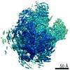
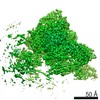
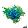
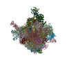
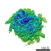
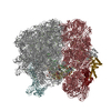
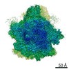
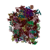
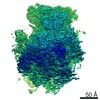
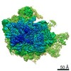
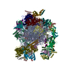
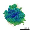
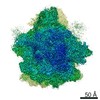
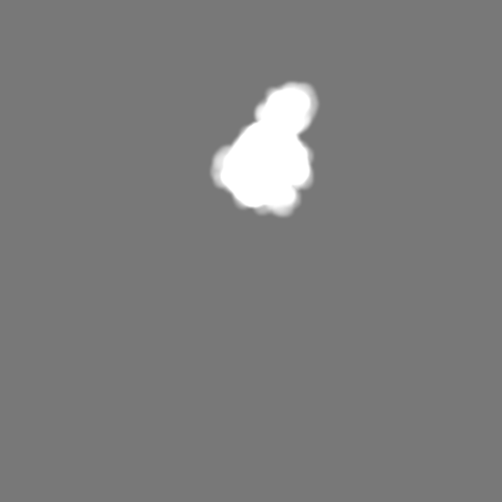
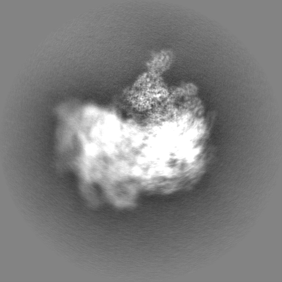
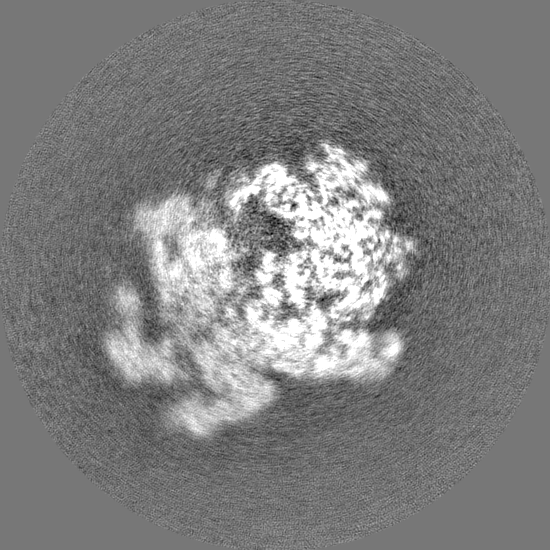
| Title | The mitochondrial ribosome from Tetrahymena thermophila, L7/L12 stalk mask | |||||||||
 Map data Map data | ||||||||||
 Sample Sample |
| |||||||||
| Biological species |  Tetrahymena thermophila SB210 (eukaryote) Tetrahymena thermophila SB210 (eukaryote) | |||||||||
| Method | single particle reconstruction / cryo EM / Resolution: 3.5 Å | |||||||||
 Authors Authors | Tobiasson V / Amunts A | |||||||||
| Funding support |  Sweden, 2 items Sweden, 2 items
| |||||||||
 Citation Citation | Journal: Elife / Year: 2020 Title: Ciliate mitoribosome illuminates evolutionary steps of mitochondrial translation. Authors: Victor Tobiasson / Alexey Amunts / Abstract: To understand the steps involved in the evolution of translation, we used , a ciliate with high coding capacity of the mitochondrial genome, as the model organism and characterized its mitochondrial ...To understand the steps involved in the evolution of translation, we used , a ciliate with high coding capacity of the mitochondrial genome, as the model organism and characterized its mitochondrial ribosome (mitoribosome) using cryo-EM. The structure of the mitoribosome reveals an assembly of 94-ribosomal proteins and four-rRNAs with an additional protein mass of ~700 kDa on the small subunit, while the large subunit lacks 5S rRNA. The structure also shows that the small subunit head is constrained, tRNA binding sites are formed by mitochondria-specific protein elements, conserved protein bS1 is excluded, and bacterial RNA polymerase binding site is blocked. We provide evidence for anintrinsic protein targeting system through visualization of mitochondria-specific mL105 by the exit tunnel that would facilitate the recruitment of a nascent polypeptide. Functional protein uS3m is encoded by three complementary genes from the nucleus and mitochondrion, establishing a link between genetic drift and mitochondrial translation. Finally, we reannotated nine open reading frames in the mitochondrial genome that code for mitoribosomal proteins. | |||||||||
| History |
|
- Structure visualization
| Movie |
 Movie viewer Movie viewer |
|---|---|
| Structure viewer | EM map: SurfViewMolmilJmol/JSmol |
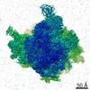
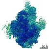
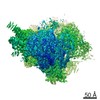
| Supplemental images |

- Downloads & links
-EMDB archive
| Map data | emd_11036.map.gz | 581 MB | EMDB map data format | |
|---|---|---|---|---|
| Header (meta data) | emd-11036-v30.xmlemd-11036.xml | 38.4 KB 38.4 KB | Display Display | EMDB header |
| Images | 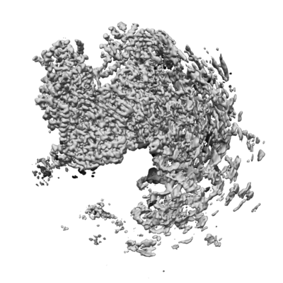 emd_11036.png emd_11036.png | 168.1 KB | ||
| Masks | emd_11036_msk_1.map | 634.7 MB | Mask map | |
| Others | emd_11036_half_map_1.map.gzemd_11036_half_map_2.map.gz | 510.5 MB 510.8 MB | ||
| Archive directory |  http://ftp.pdbj.org/pub/emdb/structures/EMD-11036ftp://ftp.pdbj.org/pub/emdb/structures/EMD-11036 http://ftp.pdbj.org/pub/emdb/structures/EMD-11036ftp://ftp.pdbj.org/pub/emdb/structures/EMD-11036 | HTTPS FTP |
-Related structure data

-Links
| EMDB pages | EMDB (EBI/PDBe) / EMDataResource |
|---|---|
| Related items in Molecule of the Month |

-Map
| File | Download / File: emd_11036.map.gz / Format: CCP4 / Size: 634.7 MB / Type: IMAGE STORED AS FLOATING POINT NUMBER (4 BYTES) | ||||||||||||||||||||||||||||||||||||||||||||||||||||||||||||||||||||
|---|---|---|---|---|---|---|---|---|---|---|---|---|---|---|---|---|---|---|---|---|---|---|---|---|---|---|---|---|---|---|---|---|---|---|---|---|---|---|---|---|---|---|---|---|---|---|---|---|---|---|---|---|---|---|---|---|---|---|---|---|---|---|---|---|---|---|---|---|---|
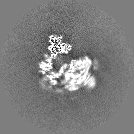
| Projections & slices | Image control
Images are generated by Spider. | ||||||||||||||||||||||||||||||||||||||||||||||||||||||||||||||||||||
| Voxel size | X=Y=Z: 1.07 Å | ||||||||||||||||||||||||||||||||||||||||||||||||||||||||||||||||||||
| Density |
| ||||||||||||||||||||||||||||||||||||||||||||||||||||||||||||||||||||
| Symmetry | Space group: 1 | ||||||||||||||||||||||||||||||||||||||||||||||||||||||||||||||||||||
| Details | EMDB XML:
CCP4 map header:
| ||||||||||||||||||||||||||||||||||||||||||||||||||||||||||||||||||||
 Z (Sec.)
 Y (Row.)
 X (Col.)


-Supplemental data
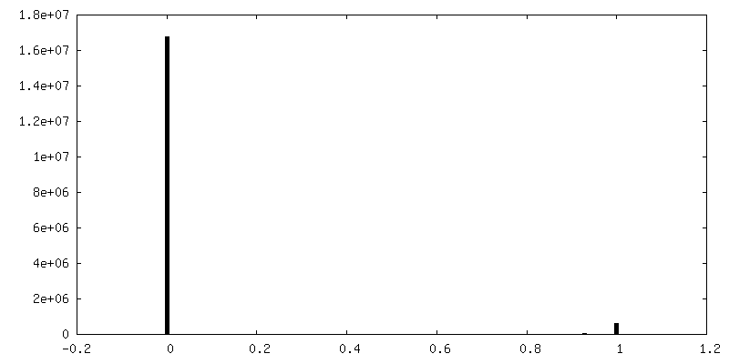
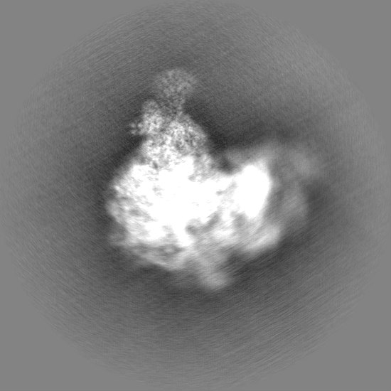
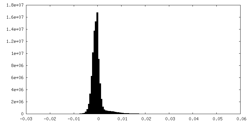
-Mask #1
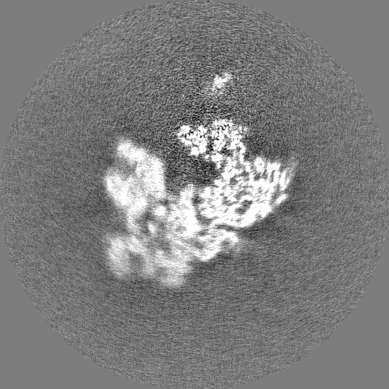
| File | emd_11036_msk_1.map | ||||||||||||
|---|---|---|---|---|---|---|---|---|---|---|---|---|---|
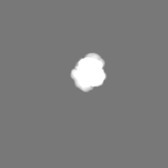
| Projections & Slices |
| ||||||||||||
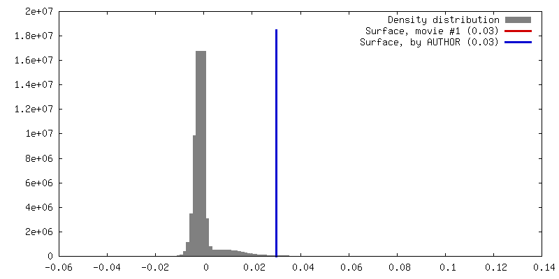
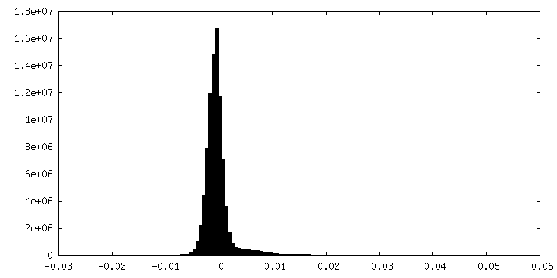
| Density Histograms |


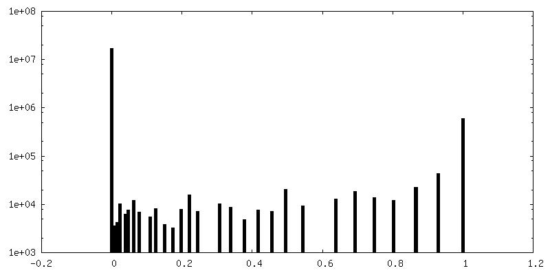
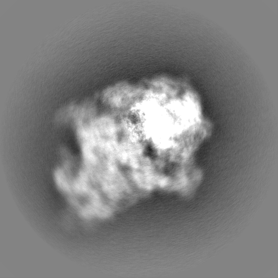
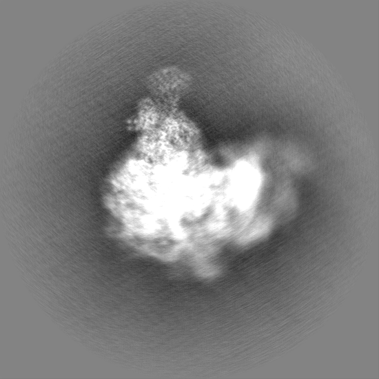
-Half map: #2
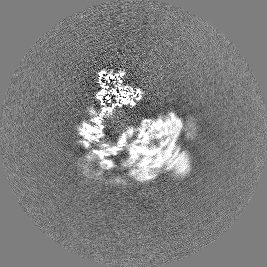
| File | emd_11036_half_map_1.map | ||||||||||||
|---|---|---|---|---|---|---|---|---|---|---|---|---|---|
| Projections & Slices |
| ||||||||||||
| Density Histograms |


-Half map: #1
| File | emd_11036_half_map_2.map | ||||||||||||
|---|---|---|---|---|---|---|---|---|---|---|---|---|---|
| Projections & Slices |
| ||||||||||||
| Density Histograms |


- Sample components
-Entire : The mitochondrial ribosome from Tetrahymena thermophila
| Entire | Name: The mitochondrial ribosome from Tetrahymena thermophila |
|---|---|
| Components |
|
-Supramolecule #1: The mitochondrial ribosome from Tetrahymena thermophila
| Supramolecule | Name: The mitochondrial ribosome from Tetrahymena thermophila type: complex / ID: 1 / Parent: 0 / Macromolecule list: #1-#94 |
|---|---|
| Source (natural) | Organism: Tetrahymena thermophila SB210 (eukaryote) |
-Supramolecule #2: Mitochondrial ribosomal large subunit from Tetrahymena thermophila
| Supramolecule | Name: Mitochondrial ribosomal large subunit from Tetrahymena thermophila type: complex / ID: 2 / Parent: 1 / Macromolecule list: #1-#48 |
|---|---|
| Source (natural) | Organism: Tetrahymena thermophila SB210 (eukaryote) |
-Supramolecule #3: Mitochondrial ribosomal small subunit from Tetrahymena thermophila
| Supramolecule | Name: Mitochondrial ribosomal small subunit from Tetrahymena thermophila type: complex / ID: 3 / Parent: 1 / Macromolecule list: #49-#94 |
|---|---|
| Source (natural) | Organism: Tetrahymena thermophila SB210 (eukaryote) |
-Experimental details
-Structure determination
| Method | cryo EM |
|---|---|
 Processing Processing | single particle reconstruction |
| Aggregation state | particle |
-Sample preparation
| Buffer | pH: 7.45 |
|---|---|
| Grid | Model: Quantifoil R2/2 / Material: COPPER / Mesh: 400 / Support film - Material: CARBON / Support film - topology: CONTINUOUS / Support film - Film thickness: 3.0 nm |
| Vitrification | Cryogen name: ETHANE |
- Electron microscopy
| Microscope | FEI TITAN KRIOS |
|---|---|
| Image recording | Film or detector model: GATAN K2 SUMMIT (4k x 4k) / Detector mode: COUNTING / Average electron dose: 30.0 e/Å2 |
| Electron beam | Acceleration voltage: 300 kV / Electron source:  FIELD EMISSION GUN FIELD EMISSION GUN |
| Electron optics | Illumination mode: FLOOD BEAM / Imaging mode: BRIGHT FIELD |
| Experimental equipment |  Model: Titan Krios / Image courtesy: FEI Company |
-Image processing
| Final reconstruction | Resolution.type: BY AUTHOR / Resolution: 3.5 Å / Resolution method: FSC 0.143 CUT-OFF / Software - Name: RELION (ver. 3.1) / Number images used: 99300 |
|---|---|
| Initial angle assignment | Type: RANDOM ASSIGNMENT |
| Final angle assignment | Type: MAXIMUM LIKELIHOOD |
