 Movie
Movie Controller


+ Open data

- Basic information
| Entry | Database: EMDB / ID: EMD-10878 | ||||||||||||
|---|---|---|---|---|---|---|---|---|---|---|---|---|---|
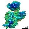
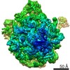
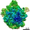
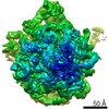
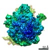
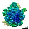
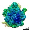
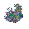
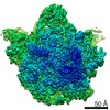
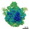
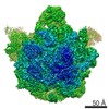
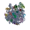
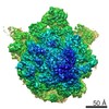
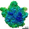
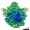
| Title | 50S ribosome subunit prepared by blotting | ||||||||||||
 Map data Map data | unsharpened final map | ||||||||||||
 Sample Sample |
| ||||||||||||
| Biological species |  | ||||||||||||
| Method | single particle reconstruction / cryo EM / Resolution: 4.4 Å | ||||||||||||
 Authors Authors | Klebl DP / Gravett MSC / Darrow M / Thompson RF / Muench SP | ||||||||||||
| Funding support |  United Kingdom, 3 items United Kingdom, 3 items
| ||||||||||||
 Citation Citation | Journal: Structure / Year: 2020 Title: Need for Speed: Examining Protein Behavior during CryoEM Grid Preparation at Different Timescales. Authors: David P Klebl / Molly S C Gravett / Dimitrios Kontziampasis / David J Wright / Robin S Bon / Diana C F Monteiro / Martin Trebbin / Frank Sobott / Howard D White / Michele C Darrow / Rebecca ...Authors: David P Klebl / Molly S C Gravett / Dimitrios Kontziampasis / David J Wright / Robin S Bon / Diana C F Monteiro / Martin Trebbin / Frank Sobott / Howard D White / Michele C Darrow / Rebecca F Thompson / Stephen P Muench /   Abstract: A host of new technologies are under development to improve the quality and reproducibility of cryoelectron microscopy (cryoEM) grid preparation. Here we have systematically investigated the ...A host of new technologies are under development to improve the quality and reproducibility of cryoelectron microscopy (cryoEM) grid preparation. Here we have systematically investigated the preparation of three macromolecular complexes using three different vitrification devices (Vitrobot, chameleon, and a time-resolved cryoEM device) on various timescales, including grids made within 6 ms (the fastest reported to date), to interrogate particle behavior at the air-water interface for different timepoints. Results demonstrate that different macromolecular complexes can respond to the thin-film environment formed during cryoEM sample preparation in highly variable ways, shedding light on why cryoEM sample preparation can be difficult to optimize. We demonstrate that reducing time between sample application and vitrification is just one tool to improve cryoEM grid quality, but that it is unlikely to be a generic "silver bullet" for improving the quality of every cryoEM sample preparation. | ||||||||||||
| History |
|
- Structure visualization
| Movie |
 Movie viewer Movie viewer |
|---|---|
| Structure viewer | EM map: SurfViewMolmilJmol/JSmol |
| Supplemental images |

- Downloads & links
-EMDB archive
| Map data | emd_10878.map.gz | 91.1 MB | EMDB map data format | |
|---|---|---|---|---|
| Header (meta data) | emd-10878-v30.xmlemd-10878.xml | 13.8 KB 13.8 KB | Display Display | EMDB header |
| FSC (resolution estimation) | emd_10878_fsc.xml | 10.8 KB | Display | FSC data file |
| Images | 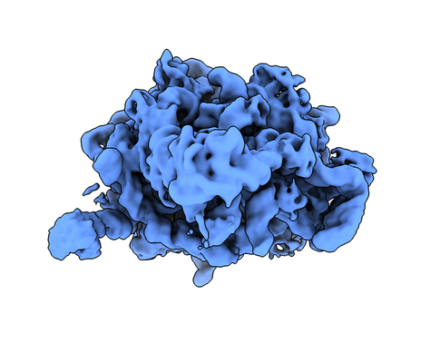 emd_10878.png emd_10878.png | 119.9 KB | ||
| Masks | emd_10878_msk_1.map | 103 MB | Mask map | |
| Others | emd_10878_half_map_1.map.gzemd_10878_half_map_2.map.gz | 80 MB 80 MB | ||
| Archive directory |  http://ftp.pdbj.org/pub/emdb/structures/EMD-10878ftp://ftp.pdbj.org/pub/emdb/structures/EMD-10878 http://ftp.pdbj.org/pub/emdb/structures/EMD-10878ftp://ftp.pdbj.org/pub/emdb/structures/EMD-10878 | HTTPS FTP |
-Validation report
| Summary document | emd_10878_validation.pdf.gz | 408.2 KB | Display | EMDB validaton report |
|---|---|---|---|---|
| Full document | emd_10878_full_validation.pdf.gz | 407.4 KB | Display | |
| Data in XML | emd_10878_validation.xml.gz | 16.8 KB | Display | |
| Arichive directory | https://ftp.pdbj.org/pub/emdb/validation_reports/EMD-10878ftp://ftp.pdbj.org/pub/emdb/validation_reports/EMD-10878 | HTTPS FTP |
-Related structure data
| Related structure data | C: citing same article ( |
|---|---|
| Similar structure data |


-Links
| EMDB pages | EMDB (EBI/PDBe) / EMDataResource |
|---|---|
| Related items in Molecule of the Month |

-Map
| File | Download / File: emd_10878.map.gz / Format: CCP4 / Size: 103 MB / Type: IMAGE STORED AS FLOATING POINT NUMBER (4 BYTES) | ||||||||||||||||||||||||||||||||||||||||||||||||||||||||||||
|---|---|---|---|---|---|---|---|---|---|---|---|---|---|---|---|---|---|---|---|---|---|---|---|---|---|---|---|---|---|---|---|---|---|---|---|---|---|---|---|---|---|---|---|---|---|---|---|---|---|---|---|---|---|---|---|---|---|---|---|---|---|
| Annotation | unsharpened final map | ||||||||||||||||||||||||||||||||||||||||||||||||||||||||||||
| Projections & slices | Image control
Images are generated by Spider. | ||||||||||||||||||||||||||||||||||||||||||||||||||||||||||||
| Voxel size | X=Y=Z: 1.065 Å | ||||||||||||||||||||||||||||||||||||||||||||||||||||||||||||
| Density |
| ||||||||||||||||||||||||||||||||||||||||||||||||||||||||||||
| Symmetry | Space group: 1 | ||||||||||||||||||||||||||||||||||||||||||||||||||||||||||||
| Details | EMDB XML:
CCP4 map header:
| ||||||||||||||||||||||||||||||||||||||||||||||||||||||||||||
 Z (Sec.)
 Y (Row.)
 X (Col.)


-Supplemental data
-Mask #1
| File | emd_10878_msk_1.map | ||||||||||||
|---|---|---|---|---|---|---|---|---|---|---|---|---|---|
| Projections & Slices |
| ||||||||||||
| Density Histograms |


-Half map: #2
| File | emd_10878_half_map_1.map | ||||||||||||
|---|---|---|---|---|---|---|---|---|---|---|---|---|---|
| Projections & Slices |
| ||||||||||||
| Density Histograms |


-Half map: #1
| File | emd_10878_half_map_2.map | ||||||||||||
|---|---|---|---|---|---|---|---|---|---|---|---|---|---|
| Projections & Slices |
| ||||||||||||
| Density Histograms |


- Sample components
-Entire : 50S ribosome from E. coli
| Entire | Name: 50S ribosome from E. coli |
|---|---|
| Components |
|
-Supramolecule #1: 50S ribosome from E. coli
| Supramolecule | Name: 50S ribosome from E. coli / type: complex / ID: 1 / Parent: 0 |
|---|---|
| Source (natural) | Organism: |
-Experimental details
-Structure determination
| Method | cryo EM |
|---|---|
 Processing Processing | single particle reconstruction |
| Aggregation state | particle |
-Sample preparation
| Concentration | 1.1 mg/mL |
|---|---|
| Buffer | pH: 7.5 |
| Vitrification | Cryogen name: ETHANE / Chamber humidity: 90 % / Chamber temperature: 293 K / Instrument: FEI VITROBOT MARK IV / Details: blot time 6 s blot force 6. |
| Details | the sample was a mixture of 30S, 50S and 70S ribosomes, purchased from New England Biolabs (P0763S) |
- Electron microscopy
| Microscope | FEI TITAN KRIOS |
|---|---|
| Image recording | Film or detector model: FEI FALCON III (4k x 4k) / Detector mode: INTEGRATING / Average exposure time: 1.5 sec. / Average electron dose: 78.0 e/Å2 |
| Electron beam | Acceleration voltage: 300 kV / Electron source:  FIELD EMISSION GUN FIELD EMISSION GUN |
| Electron optics | Illumination mode: SPOT SCAN / Imaging mode: BRIGHT FIELD |
| Experimental equipment |  Model: Titan Krios / Image courtesy: FEI Company |
-Image processing
| Final reconstruction | Applied symmetry - Point group: C1 (asymmetric) / Resolution.type: BY AUTHOR / Resolution: 4.4 Å / Resolution method: FSC 0.143 CUT-OFF / Software - Name: RELION / Number images used: 84671 |
|---|---|
| Initial angle assignment | Type: NOT APPLICABLE |
| Final angle assignment | Type: MAXIMUM LIKELIHOOD / Software - Name: RELION Details: The final angular assignment was done in a bigger, combined dataset which was then split into the original datasets to generate this reconstruction |
| FSC plot (resolution estimation) |  |
