Movie
Movie Controller

[English] 日本語
 Yorodumi
- EMDB-1081: Seeing GroEL at 6 A resolution by single particle electron cryomi... -
+ Open data

- Basic information
| Entry | Database: EMDB / ID: EMD-1081 | |||||||||
|---|---|---|---|---|---|---|---|---|---|---|
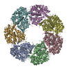
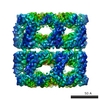
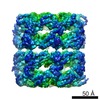
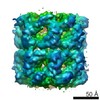
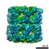
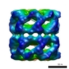
| Title | Seeing GroEL at 6 A resolution by single particle electron cryomicroscopy. | |||||||||
 Map data Map data | This is the density map for the final published ~6 A resolution GroEL structure. | |||||||||
 Sample Sample |
| |||||||||
| Biological species |  | |||||||||
| Method | single particle reconstruction / cryo EM / Resolution: 6.0 Å | |||||||||
 Authors Authors | Ludtke SJ / Chen D / Song J / Chuang DT / Chiu W | |||||||||
 Citation Citation | Journal: Structure / Year: 2004 Title: Seeing GroEL at 6 A resolution by single particle electron cryomicroscopy. Authors: Steven J Ludtke / Dong-Hua Chen / Jiu-Li Song / David T Chuang / Wah Chiu /  Abstract: We present a reconstruction of native GroEL by electron cryomicroscopy (cryo-EM) and single particle analysis at 6 A resolution. alpha helices are clearly visible and beta sheet density is also ...We present a reconstruction of native GroEL by electron cryomicroscopy (cryo-EM) and single particle analysis at 6 A resolution. alpha helices are clearly visible and beta sheet density is also visible at this resolution. While the overall conformation of this structure is quite consistent with the published X-ray data, a measurable shift in the positions of three alpha helices in the intermediate domain is observed, not consistent with any of the 7 monomeric structures in the Protein Data Bank model (1OEL). In addition, there is evidence for slight rearrangement or flexibility in parts of the apical domain. The 6 A resolution cryo-EM GroEL structure clearly demonstrates the veracity and expanding scope of cryo-EM and the single particle reconstruction technique for macromolecular machines. | |||||||||
| History |
|
- Structure visualization
| Movie |
 Movie viewer Movie viewer |
|---|---|
| Structure viewer | EM map: SurfViewMolmilJmol/JSmol |
| Supplemental images |
- Downloads & links
-EMDB archive
| Map data | emd_1081.map.gz | 7.4 MB | EMDB map data format | |
|---|---|---|---|---|
| Header (meta data) | emd-1081-v30.xmlemd-1081.xml | 9.1 KB 9.1 KB | Display Display | EMDB header |
| Images |  1081.gif 1081.gif | 34.4 KB | ||
| Archive directory |  http://ftp.pdbj.org/pub/emdb/structures/EMD-1081ftp://ftp.pdbj.org/pub/emdb/structures/EMD-1081 http://ftp.pdbj.org/pub/emdb/structures/EMD-1081ftp://ftp.pdbj.org/pub/emdb/structures/EMD-1081 | HTTPS FTP |
-Related structure data
| Similar structure data |
|---|


-Links
| EMDB pages | EMDB (EBI/PDBe) / EMDataResource |
|---|
-Map
| File | Download / File: emd_1081.map.gz / Format: CCP4 / Size: 7.8 MB / Type: IMAGE STORED AS FLOATING POINT NUMBER (4 BYTES) | ||||||||||||||||||||||||||||||||||||||||||||||||||||||||||||||||||||
|---|---|---|---|---|---|---|---|---|---|---|---|---|---|---|---|---|---|---|---|---|---|---|---|---|---|---|---|---|---|---|---|---|---|---|---|---|---|---|---|---|---|---|---|---|---|---|---|---|---|---|---|---|---|---|---|---|---|---|---|---|---|---|---|---|---|---|---|---|---|
| Annotation | This is the density map for the final published ~6 A resolution GroEL structure. | ||||||||||||||||||||||||||||||||||||||||||||||||||||||||||||||||||||
| Projections & slices | Image control
Images are generated by Spider. | ||||||||||||||||||||||||||||||||||||||||||||||||||||||||||||||||||||
| Voxel size | X=Y=Z: 2.08 Å | ||||||||||||||||||||||||||||||||||||||||||||||||||||||||||||||||||||
| Density |
| ||||||||||||||||||||||||||||||||||||||||||||||||||||||||||||||||||||
| Symmetry | Space group: 1 | ||||||||||||||||||||||||||||||||||||||||||||||||||||||||||||||||||||
| Details | EMDB XML:
CCP4 map header:
| ||||||||||||||||||||||||||||||||||||||||||||||||||||||||||||||||||||
 Z (Sec.)
 Y (Row.)
 X (Col.)


-Supplemental data
- Sample components
-Entire : native naked GroEL
| Entire | Name: native naked GroEL |
|---|---|
| Components |
|
-Supramolecule #1000: native naked GroEL
| Supramolecule | Name: native naked GroEL / type: sample / ID: 1000 / Oligomeric state: homo 14-mer / Number unique components: 1 |
|---|---|
| Molecular weight | Theoretical: 800 KDa |
-Macromolecule #1: GroEL
| Macromolecule | Name: GroEL / type: protein_or_peptide / ID: 1 / Number of copies: 14 / Oligomeric state: 14mer / Recombinant expression: Yes |
|---|---|
| Source (natural) | Organism: |
| Molecular weight | Experimental: 57 KDa |
| Recombinant expression | Organism: ESts CG-712 / Recombinant plasmid: pGroESL |
-Experimental details
-Structure determination
| Method | cryo EM |
|---|---|
 Processing Processing | single particle reconstruction |
| Aggregation state | particle |
-Sample preparation
| Concentration | 1.0 mg/mL |
|---|---|
| Vitrification | Cryogen name: ETHANE / Instrument: HOMEMADE PLUNGER / Details: Vitrification instrument: Manual gravity plunger |
- Electron microscopy
| Microscope | JEOL 2010F |
|---|---|
| Image recording | Category: FILM / Film or detector model: KODAK SO-163 FILM / Digitization - Scanner: ZEISS SCAI / Digitization - Sampling interval: 7 µm / Number real images: 42 / Average electron dose: 18 e/Å2 Details: Data was median filtered to 10.5 microns/pixel after scanning. Bits/pixel: 8 |
| Electron beam | Acceleration voltage: 200 kV / Electron source:  FIELD EMISSION GUN FIELD EMISSION GUN |
| Electron optics | Illumination mode: FLOOD BEAM / Imaging mode: BRIGHT FIELD / Cs: 1.0 mm / Nominal defocus max: 2.56 µm / Nominal defocus min: 0.794 µm / Nominal magnification: 50000 |
| Sample stage | Specimen holder: Side entry / Specimen holder model: GATAN LIQUID NITROGEN |
-Image processing
| Details | Note that class averages are EMAN-style reference-based, not MSA. |
|---|---|
| CTF correction | Details: per-particle phase flipping pre-processing, amplitude correction during class averaging |
| Final reconstruction | Applied symmetry - Point group: D7 (2x7 fold dihedral) / Resolution.type: BY AUTHOR / Resolution: 6.0 Å / Resolution method: FSC 0.5 CUT-OFF / Software - Name: EMAN / Number images used: 39085 |
| Final two d classification | Number classes: 295 |
-Atomic model buiding 1
| Initial model | PDB ID: |
|---|---|
| Software | Name: foldhunter, EMAN |
| Details | Protocol: Rigid Body. rigid body fitting of 1 monomer from 1OEL initially, followed by rigid body refinement of each domain, apical, intermediate and equatorial. |
| Refinement | Space: REAL / Protocol: RIGID BODY FIT / Target criteria: correlation coefficient |