Movie
Movie Controller

[English] 日本語
 Yorodumi
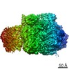
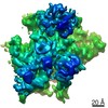
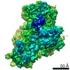
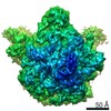
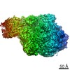
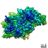
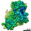
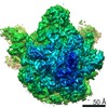
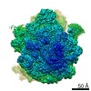
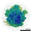
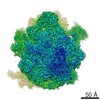
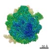
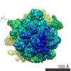
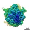
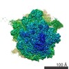
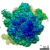
- EMDB-10683: 70S ribosome from chloramphenicol-treated Mycoplasma pneumoniae cells -
+ Open data

- Basic information
| Entry | Database: EMDB / ID: EMD-10683 | |||||||||
|---|---|---|---|---|---|---|---|---|---|---|
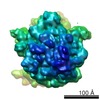
| Title | 70S ribosome from chloramphenicol-treated Mycoplasma pneumoniae cells | |||||||||
 Map data Map data | in-cell 70S ribosome from Mycoplasma pneumoniae cells after chloramphenicol treatment for 15 minutes | |||||||||
 Sample Sample |
| |||||||||
| Biological species |  Mycoplasma pneumoniae M129 (bacteria) Mycoplasma pneumoniae M129 (bacteria) | |||||||||
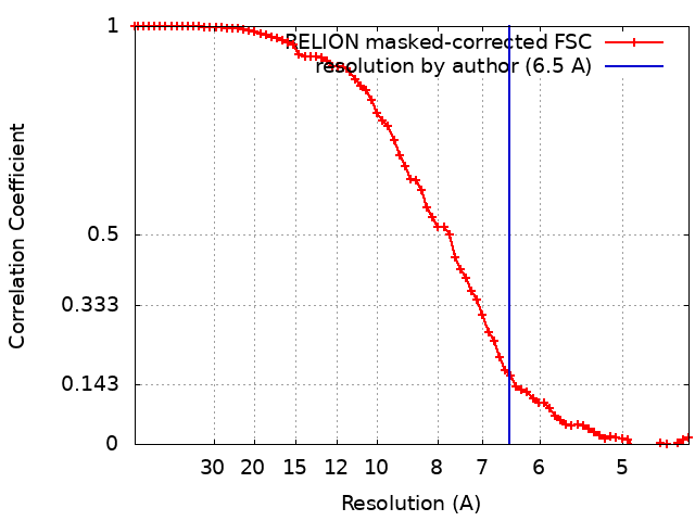
| Method | subtomogram averaging / cryo EM / Resolution: 6.5 Å | |||||||||
 Authors Authors | Mahamid J / Xue L | |||||||||
| Funding support |  Germany, 1 items Germany, 1 items
| |||||||||
 Citation Citation | Journal: Science / Year: 2020 Title: In-cell architecture of an actively transcribing-translating expressome. Authors: Francis J O'Reilly / Liang Xue / Andrea Graziadei / Ludwig Sinn / Swantje Lenz / Dimitry Tegunov / Cedric Blötz / Neil Singh / Wim J H Hagen / Patrick Cramer / Jörg Stülke / Julia Mahamid ...Authors: Francis J O'Reilly / Liang Xue / Andrea Graziadei / Ludwig Sinn / Swantje Lenz / Dimitry Tegunov / Cedric Blötz / Neil Singh / Wim J H Hagen / Patrick Cramer / Jörg Stülke / Julia Mahamid / Juri Rappsilber /  Abstract: Structural biology studies performed inside cells can capture molecular machines in action within their native context. In this work, we developed an integrative in-cell structural approach using the ...Structural biology studies performed inside cells can capture molecular machines in action within their native context. In this work, we developed an integrative in-cell structural approach using the genome-reduced human pathogen We combined whole-cell cross-linking mass spectrometry, cellular cryo-electron tomography, and integrative modeling to determine an in-cell architecture of a transcribing and translating expressome at subnanometer resolution. The expressome comprises RNA polymerase (RNAP), the ribosome, and the transcription elongation factors NusG and NusA. We pinpointed NusA at the interface between a NusG-bound elongating RNAP and the ribosome and propose that it can mediate transcription-translation coupling. Translation inhibition dissociated the expressome, whereas transcription inhibition stalled and rearranged it. Thus, the active expressome architecture requires both translation and transcription elongation within the cell. | |||||||||
| History |
|
- Structure visualization
| Movie |
 Movie viewer Movie viewer |
|---|---|
| Structure viewer | EM map: SurfViewMolmilJmol/JSmol |
| Supplemental images |
- Downloads & links
-EMDB archive
| Map data | emd_10683.map.gz | 3.8 MB | EMDB map data format | |
|---|---|---|---|---|
| Header (meta data) | emd-10683-v30.xmlemd-10683.xml | 15 KB 15 KB | Display Display | EMDB header |
| FSC (resolution estimation) | emd_10683_fsc.xml | 7.1 KB | Display | FSC data file |
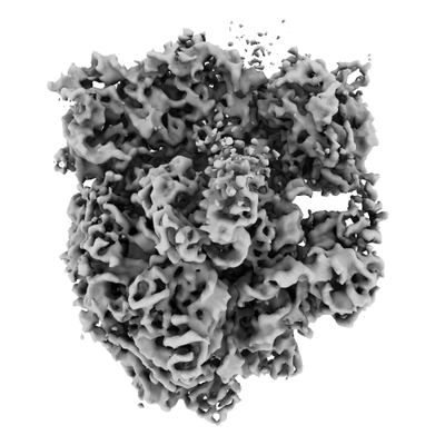
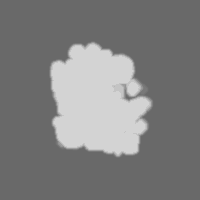
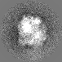
| Images |  emd_10683.png emd_10683.png | 79.1 KB | ||
| Masks | emd_10683_msk_1.map | 30.5 MB | Mask map | |
| Others | emd_10683_half_map_1.map.gzemd_10683_half_map_2.map.gz | 23.4 MB 23.4 MB | ||
| Archive directory |  http://ftp.pdbj.org/pub/emdb/structures/EMD-10683ftp://ftp.pdbj.org/pub/emdb/structures/EMD-10683 http://ftp.pdbj.org/pub/emdb/structures/EMD-10683ftp://ftp.pdbj.org/pub/emdb/structures/EMD-10683 | HTTPS FTP |
-Related structure data
| Related structure data | C: citing same article ( |
|---|---|
| Similar structure data |
-Links
| EMDB pages | EMDB (EBI/PDBe) / EMDataResource |
|---|---|
| Related items in Molecule of the Month |

-Map
| File | Download / File: emd_10683.map.gz / Format: CCP4 / Size: 30.5 MB / Type: IMAGE STORED AS FLOATING POINT NUMBER (4 BYTES) | ||||||||||||||||||||||||||||||||||||||||||||||||||||||||||||
|---|---|---|---|---|---|---|---|---|---|---|---|---|---|---|---|---|---|---|---|---|---|---|---|---|---|---|---|---|---|---|---|---|---|---|---|---|---|---|---|---|---|---|---|---|---|---|---|---|---|---|---|---|---|---|---|---|---|---|---|---|---|
| Annotation | in-cell 70S ribosome from Mycoplasma pneumoniae cells after chloramphenicol treatment for 15 minutes | ||||||||||||||||||||||||||||||||||||||||||||||||||||||||||||
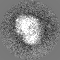
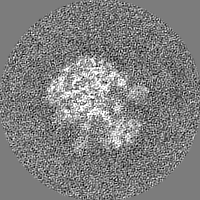
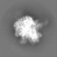
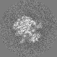
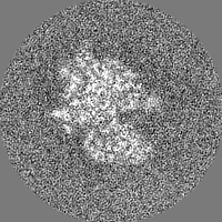
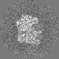
| Projections & slices | Image control
Images are generated by Spider. | ||||||||||||||||||||||||||||||||||||||||||||||||||||||||||||
| Voxel size | X=Y=Z: 2.2 Å | ||||||||||||||||||||||||||||||||||||||||||||||||||||||||||||
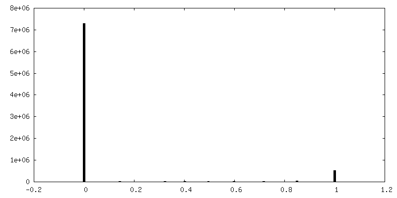
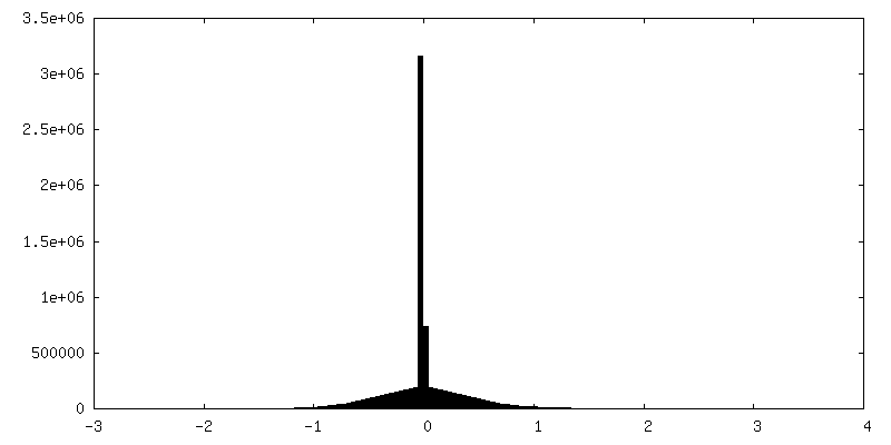
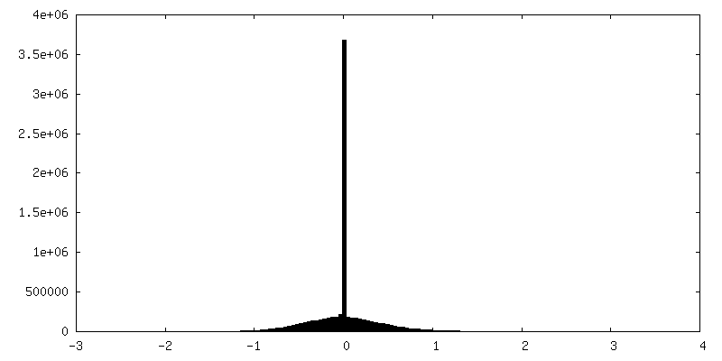
| Density |
| ||||||||||||||||||||||||||||||||||||||||||||||||||||||||||||
| Symmetry | Space group: 1 | ||||||||||||||||||||||||||||||||||||||||||||||||||||||||||||
| Details | EMDB XML:
CCP4 map header:
| ||||||||||||||||||||||||||||||||||||||||||||||||||||||||||||
 Z (Sec.)
 Y (Row.)
 X (Col.)


-Supplemental data
-Mask #1
| File | emd_10683_msk_1.map | ||||||||||||
|---|---|---|---|---|---|---|---|---|---|---|---|---|---|
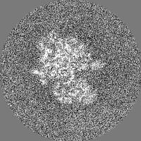
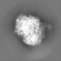
| Projections & Slices |
| ||||||||||||
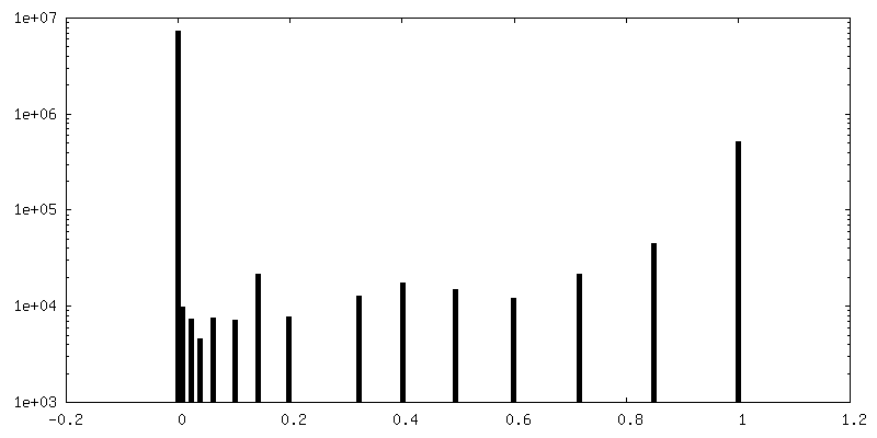
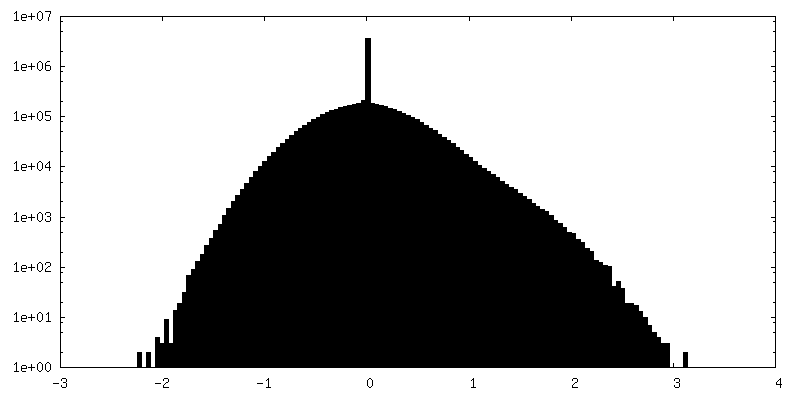
| Density Histograms |


-Half map: in-cell 70S ribosome from chloramphenicol-treated Mycoplasma pneumoniae cells,...
| File | emd_10683_half_map_1.map | ||||||||||||
|---|---|---|---|---|---|---|---|---|---|---|---|---|---|
| Annotation | in-cell 70S ribosome from chloramphenicol-treated Mycoplasma pneumoniae cells, half map 1 | ||||||||||||
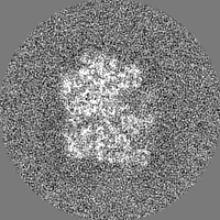
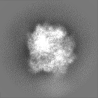
| Projections & Slices |
| ||||||||||||
| Density Histograms |


-Half map: in-cell 70S ribosome from chloramphenicol-treated Mycoplasma pneumoniae cells,...
| File | emd_10683_half_map_2.map | ||||||||||||
|---|---|---|---|---|---|---|---|---|---|---|---|---|---|
| Annotation | in-cell 70S ribosome from chloramphenicol-treated Mycoplasma pneumoniae cells, half map 2 | ||||||||||||
| Projections & Slices |
| ||||||||||||
| Density Histograms |
- Sample components
-Entire : Chloramphenicol-treated Mycoplasma pneumoniae M129 cells
| Entire | Name: Chloramphenicol-treated Mycoplasma pneumoniae M129 cells |
|---|---|
| Components |
|
-Supramolecule #1: Chloramphenicol-treated Mycoplasma pneumoniae M129 cells
| Supramolecule | Name: Chloramphenicol-treated Mycoplasma pneumoniae M129 cells type: cell / ID: 1 / Parent: 0 Details: Wild-type Mycoplasma pneumoniae M129 cells were treated with 0.5 mg/ml chloramphenicol (Cm) for 15 minutes prior to plunge-freezing |
|---|---|
| Source (natural) | Organism: Mycoplasma pneumoniae M129 (bacteria) |
-Experimental details
-Structure determination
| Method | cryo EM |
|---|---|
 Processing Processing | subtomogram averaging |
| Aggregation state | cell |
-Sample preparation
| Buffer | pH: 7.4 Details: modified Hayflick medium as described in Halbedel, Hames, and Stulke 2004, with chloramphenicol (Cm) at a final concentration of 0.5 mg/ml |
|---|---|
| Grid | Model: Quantifoil R2/1 / Material: GOLD / Support film - Material: CARBON / Support film - topology: HOLEY / Pretreatment - Type: GLOW DISCHARGE |
| Vitrification | Cryogen name: ETHANE-PROPANE / Chamber humidity: 45 % / Instrument: HOMEMADE PLUNGER |
- Electron microscopy
| Microscope | FEI TITAN KRIOS |
|---|---|
| Image recording | Film or detector model: GATAN K2 SUMMIT (4k x 4k) / Detector mode: COUNTING / Average electron dose: 2.9 e/Å2 |
| Electron beam | Acceleration voltage: 300 kV / Electron source:  FIELD EMISSION GUN FIELD EMISSION GUN |
| Electron optics | Calibrated defocus max: 4.5 µm / Calibrated defocus min: 1.5 µm / Calibrated magnification: 81000 / Illumination mode: FLOOD BEAM / Imaging mode: BRIGHT FIELD / Cs: 2.7 mm |
| Sample stage | Specimen holder model: FEI TITAN KRIOS AUTOGRID HOLDER / Cooling holder cryogen: NITROGEN |
| Experimental equipment |  Model: Titan Krios / Image courtesy: FEI Company |