 ムービー
ムービー コントローラー


+ データを開く

- 基本情報
基本情報
| 登録情報 | データベース: EMDB / ID: EMD-1062 | |||||||||
|---|---|---|---|---|---|---|---|---|---|---|
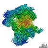
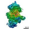
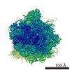
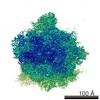
| タイトル | Three-dimensional structure of C complex spliceosomes by electron microscopy. | |||||||||
 マップデータ マップデータ | Map of C-complex of the human spliceosome. | |||||||||
 試料 試料 |
| |||||||||
| 生物種 |  Homo sapiens (ヒト) Homo sapiens (ヒト) | |||||||||
| 手法 | 単粒子再構成法 / クライオ電子顕微鏡法 / ネガティブ染色法 / 解像度: 30.0 Å | |||||||||
 データ登録者 データ登録者 | Jurica MS / Sousa D / Moore MJ / Grigorieff N | |||||||||
 引用 引用 | ジャーナル: Nat Struct Mol Biol / 年: 2004 タイトル: Three-dimensional structure of C complex spliceosomes by electron microscopy. 著者: Melissa S Jurica / Duncan Sousa / Melissa J Moore / Nikolaus Grigorieff /  要旨: The spliceosome is a multimegadalton RNA-protein machine that removes noncoding sequences from nascent pre-mRNAs. Recruitment of the spliceosome to splice sites and subsequent splicing require a ...The spliceosome is a multimegadalton RNA-protein machine that removes noncoding sequences from nascent pre-mRNAs. Recruitment of the spliceosome to splice sites and subsequent splicing require a series of dynamic interactions among the spliceosome's component U snRNPs and many additional protein factors. These dynamics present several challenges for structural analyses, including purification of stable complexes to compositional homogeneity and assessment of conformational heterogeneity. We have isolated spliceosomes arrested before the second chemical step of splicing (C complex) in which U2, U5 and U6 snRNAs are stably associated. Using electron microscopy, we obtained images of C complex spliceosomes under cryogenic conditions and determined a three-dimensional structure of a core complex to a resolution of 30 A. The structure reveals a particle of dimensions 27 x 22 x 24 nm with a relatively open arrangement of three primary domains. | |||||||||
| 履歴 |
|
- 構造の表示
構造の表示
| ムービー |
 ムービービューア ムービービューア |
|---|---|
| 構造ビューア | EMマップ: SurfViewMolmilJmol/JSmol |
| 添付画像 |
 UCSF Chimera
- ダウンロードとリンク
-EMDBアーカイブ
| マップデータ | emd_1062.map.gz | 3.3 MB | EMDBマップデータ形式 | |
|---|---|---|---|---|
| ヘッダ (付随情報) | emd-1062-v30.xmlemd-1062.xml | 9.8 KB 9.8 KB | 表示 表示 | EMDBヘッダ |
| 画像 |  1062.gif 1062.gif | 46.7 KB | ||
| アーカイブディレクトリ |  http://ftp.pdbj.org/pub/emdb/structures/EMD-1062ftp://ftp.pdbj.org/pub/emdb/structures/EMD-1062 http://ftp.pdbj.org/pub/emdb/structures/EMD-1062ftp://ftp.pdbj.org/pub/emdb/structures/EMD-1062 | HTTPS FTP |
-検証レポート
| 文書・要旨 | emd_1062_validation.pdf.gz | 188.2 KB | 表示 | EMDB検証レポート |
|---|---|---|---|---|
| 文書・詳細版 | emd_1062_full_validation.pdf.gz | 187.3 KB | 表示 | |
| XML形式データ | emd_1062_validation.xml.gz | 5.6 KB | 表示 | |
| アーカイブディレクトリ | https://ftp.pdbj.org/pub/emdb/validation_reports/EMD-1062ftp://ftp.pdbj.org/pub/emdb/validation_reports/EMD-1062 | HTTPS FTP |
-関連構造データ


-リンク
| EMDBのページ | EMDB (EBI/PDBe) / EMDataResource |
|---|
-マップ
| ファイル | ダウンロード / ファイル: emd_1062.map.gz / 形式: CCP4 / 大きさ: 7.8 MB / タイプ: IMAGE STORED AS FLOATING POINT NUMBER (4 BYTES) | ||||||||||||||||||||||||||||||||||||||||||||||||||||||||||||||||||||
|---|---|---|---|---|---|---|---|---|---|---|---|---|---|---|---|---|---|---|---|---|---|---|---|---|---|---|---|---|---|---|---|---|---|---|---|---|---|---|---|---|---|---|---|---|---|---|---|---|---|---|---|---|---|---|---|---|---|---|---|---|---|---|---|---|---|---|---|---|---|
| 注釈 | Map of C-complex of the human spliceosome. | ||||||||||||||||||||||||||||||||||||||||||||||||||||||||||||||||||||
| 投影像・断面図 | 画像のコントロール
画像は Spider により作成 | ||||||||||||||||||||||||||||||||||||||||||||||||||||||||||||||||||||
| ボクセルのサイズ | X=Y=Z: 4.6667 Å | ||||||||||||||||||||||||||||||||||||||||||||||||||||||||||||||||||||
| 密度 |
| ||||||||||||||||||||||||||||||||||||||||||||||||||||||||||||||||||||
| 対称性 | 空間群: 1 | ||||||||||||||||||||||||||||||||||||||||||||||||||||||||||||||||||||
| 詳細 | EMDB XML:
CCP4マップ ヘッダ情報:
| ||||||||||||||||||||||||||||||||||||||||||||||||||||||||||||||||||||
 Z (Sec.)
 Y (Row.)
 X (Col.)


-添付データ
- 試料の構成要素
-全体 : C-complex of the human spliceosome
| 全体 | 名称: C-complex of the human spliceosome |
|---|---|
| 要素 |
|
-超分子 #1000: C-complex of the human spliceosome
| 超分子 | 名称: C-complex of the human spliceosome / タイプ: sample / ID: 1000 / Number unique components: 1 |
|---|---|
| 分子量 | 理論値: 2.6 MDa 手法: The molecular weight of core C-complex components were added together. The map becomes discontinous at either higher or lower weights. |
-超分子 #1: C-complex Spliceosome
| 超分子 | 名称: C-complex Spliceosome / タイプ: organelle_or_cellular_component / ID: 1 / Name.synonym: Spliceosome / コピー数: 1 / 集合状態: monomer / 組換発現: No |
|---|---|
| 由来(天然) | 生物種: Homo sapiens (ヒト) / 別称: Human / 細胞: HeLa / Organelle: Nucleus / 細胞中の位置: Nucleus |
| 分子量 | 実験値: 2.6 MDa |
-実験情報
-構造解析
| 手法 | ネガティブ染色法, クライオ電子顕微鏡法 |
|---|---|
 解析 解析 | 単粒子再構成法 |
| 試料の集合状態 | particle |
-試料調製
| 緩衝液 | pH: 7.5 詳細: 150mM KCl, 5mM EDTA, 20mM Tris, 10mM maltose, and 5% glycerol |
|---|---|
| 染色 | タイプ: NEGATIVE 詳細: Thin carbon is floated for 1 minute on the sample. The carbon is transferred onto a solution of 1% uranyl formate, sample side down, and then picked up with a holey carbon grid (Quantifoil). ...詳細: Thin carbon is floated for 1 minute on the sample. The carbon is transferred onto a solution of 1% uranyl formate, sample side down, and then picked up with a holey carbon grid (Quantifoil). A second layer of thin carbon is sandwiched on top of the sample. |
| グリッド | 詳細: 400 mesh copper grid |
| 凍結 | 凍結剤: NITROGEN / 装置: HOMEMADE PLUNGER / 詳細: Vitrification instrument: human hand. none / 手法: See experimental details. |
- 電子顕微鏡法
| 顕微鏡 | FEI/PHILIPS CM12 |
|---|---|
| 温度 | 最低: 90 K / 最高: 100 K / 平均: 95 K |
| アライメント法 | Legacy - 非点収差: objective lens astigmatism was corrected at 200,000 times magnification |
| 詳細 | MICROSCOPE Philips CM12 |
| 撮影 | カテゴリ: FILM / フィルム・検出器のモデル: KODAK SO-163 FILM / デジタル化 - スキャナー: ZEISS SCAI / デジタル化 - サンプリング間隔: 7 µm / 実像数: 72 / 平均電子線量: 10 e/Å2 / ビット/ピクセル: 8 |
| 電子線 | 加速電圧: 120 kV / 電子線源: LAB6 |
| 電子光学系 | 倍率(補正後): 60000 / 照射モード: FLOOD BEAM / 撮影モード: BRIGHT FIELD / Cs: 2 mm / 最大 デフォーカス(公称値): 20.0 µm / 最小 デフォーカス(公称値): 1.0 µm / 倍率(公称値): 60000 |
| 試料ステージ | 試料ホルダー: Side entry liquid nitrogen-cooled cryo specimen holder. 試料ホルダーモデル: OTHER / Tilt angle min: 35 / Tilt angle max: 45 |
-画像解析
| 詳細 | Image tilt pairs were collected manually and processed using Spider and Frealign. |
|---|---|
| CTF補正 | 詳細: CTF correction of each particle |
| 最終 再構成 | 想定した対称性 - 点群: C1 (非対称) / アルゴリズム: OTHER / 解像度のタイプ: BY AUTHOR / 解像度: 30.0 Å / 解像度の算出法: FSC 0.5 CUT-OFF / ソフトウェア - 名称: Spider and Frealign 詳細: The final map was calculated from one dataset and filtered to 20 angstroms resolution. 使用した粒子像数: 1834 |
