- EMDB-1057: The structure of echovirus type 12 bound to a two-domain fragment... -
+
データを開く
IDまたはキーワード:
読み込み中...
-
基本情報
登録情報
データベース: EMDB / ID: EMD-1057
タイトル
The structure of echovirus type 12 bound to a two-domain fragment of its cellular attachment protein decay-accelerating factor (CD 55).
マップデータ
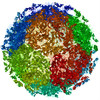
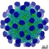
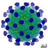
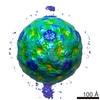
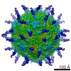
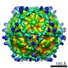
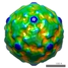
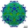
Three dimensional reconstruction of echovirus type 12 bound to domains three and four of its cellular receptor decay-accelerating factor. Calculated from cryo-negative stain images
試料
試料: Echovirus type 12 bound to decay accelerating factor domains 3 and 4
ウイルス: Human echovirus 12 (ウイルス)
細胞器官・細胞要素: Decay accelerating factor domains 3 and 4
機能・相同性
機能・相同性情報
regulation of lipopolysaccharide-mediated signaling pathway / negative regulation of complement activation / regulation of complement-dependent cytotoxicity / regulation of complement activation / respiratory burst / positive regulation of CD4-positive, alpha-beta T cell activation / positive regulation of CD4-positive, alpha-beta T cell proliferation / Class B/2 (Secretin family receptors) / symbiont-mediated suppression of host cytoplasmic pattern recognition receptor signaling pathway via inhibition of RIG-I activity / ficolin-1-rich granule membrane ...regulation of lipopolysaccharide-mediated signaling pathway / negative regulation of complement activation / regulation of complement-dependent cytotoxicity / regulation of complement activation / respiratory burst / positive regulation of CD4-positive, alpha-beta T cell activation / positive regulation of CD4-positive, alpha-beta T cell proliferation / Class B/2 (Secretin family receptors) / symbiont-mediated suppression of host cytoplasmic pattern recognition receptor signaling pathway via inhibition of RIG-I activity / ficolin-1-rich granule membrane / complement activation, classical pathway / transport vesicle / side of membrane / COPI-mediated anterograde transport / endoplasmic reticulum-Golgi intermediate compartment membrane / secretory granule membrane / Regulation of Complement cascade / picornain 2A / symbiont-mediated suppression of host mRNA export from nucleus / symbiont genome entry into host cell via pore formation in plasma membrane / picornain 3C / T=pseudo3 icosahedral viral capsid / positive regulation of T cell cytokine production / host cell cytoplasmic vesicle membrane / nucleoside-triphosphate phosphatase / channel activity / positive regulation of cytosolic calcium ion concentration / virus receptor activity / monoatomic ion transmembrane transport / DNA replication / RNA helicase activity / membrane raft / endocytosis involved in viral entry into host cell / Golgi membrane / symbiont-mediated activation of host autophagy / innate immune response / RNA-directed RNA polymerase / cysteine-type endopeptidase activity / viral RNA genome replication / RNA-directed RNA polymerase activity / DNA-templated transcription / lipid binding / Neutrophil degranulation / virion attachment to host cell / host cell nucleus / structural molecule activity / cell surface / ATP hydrolysis activity / proteolysis / RNA binding / extracellular exosome / extracellular region / zinc ion binding / ATP binding / membrane / plasma membrane 類似検索 - 分子機能
: / Sushi repeat (SCR repeat) / Domain abundant in complement control proteins; SUSHI repeat; short complement-like repeat (SCR) / Picornavirus coat protein VP4 superfamily / Sushi/SCR/CCP domain / Sushi/CCP/SCR domain profile. / Sushi/SCR/CCP superfamily / Poliovirus 3A protein-like / Poliovirus 3A protein like / Picornavirus 2B protein ...: / Sushi repeat (SCR repeat) / Domain abundant in complement control proteins; SUSHI repeat; short complement-like repeat (SCR) / Picornavirus coat protein VP4 superfamily / Sushi/SCR/CCP domain / Sushi/CCP/SCR domain profile. / Sushi/SCR/CCP superfamily / Poliovirus 3A protein-like / Poliovirus 3A protein like / Picornavirus 2B protein / Poliovirus core protein 3a, soluble domain / Picornavirus 2B protein / Peptidase C3, picornavirus core protein 2A / Picornavirus core protein 2A / Picornavirus coat protein VP4 / Picornavirus coat protein (VP4) / Peptidase C3A/C3B, picornaviral / 3C cysteine protease (picornain 3C) / Picornavirales 3C/3C-like protease domain / Picornavirales 3C/3C-like protease domain profile. / Picornavirus capsid / picornavirus capsid protein / Helicase, superfamily 3, single-stranded RNA virus / Superfamily 3 helicase of positive ssRNA viruses domain profile. / Helicase, superfamily 3, single-stranded DNA/RNA virus / RNA helicase / Picornavirus/Calicivirus coat protein / Viral coat protein subunit / RNA-directed RNA polymerase, C-terminal domain / Viral RNA-dependent RNA polymerase / Reverse transcriptase/Diguanylate cyclase domain / RNA-directed RNA polymerase, catalytic domain / RdRp of positive ssRNA viruses catalytic domain profile. / ATPases associated with a variety of cellular activities / AAA+ ATPase domain / Peptidase S1, PA clan, chymotrypsin-like fold / Peptidase S1, PA clan / DNA/RNA polymerase superfamily / P-loop containing nucleoside triphosphate hydrolase 類似検索 - ドメイン・相同性
ジャーナル: J Biol Chem / 年: 2004 タイトル: The structure of echovirus type 12 bound to a two-domain fragment of its cellular attachment protein decay-accelerating factor (CD 55). 著者: David Bhella / Ian G Goodfellow / Pietro Roversi / David Pettigrew / Yasmin Chaudhry / David J Evans / Susan M Lea / 要旨: Echovirus type 12 (EV12), an Enterovirus of the Picornaviridae family, uses the complement regulator decay-accelerating factor (DAF, CD55) as a cellular receptor. We have calculated a three- ...Echovirus type 12 (EV12), an Enterovirus of the Picornaviridae family, uses the complement regulator decay-accelerating factor (DAF, CD55) as a cellular receptor. We have calculated a three-dimensional reconstruction of EV12 bound to a fragment of DAF consisting of short consensus repeat domains 3 and 4 from cryo-negative stain electron microscopy data (EMD code 1057). This shows that, as for an earlier reconstruction of the related echovirus type 7 bound to DAF, attachment is not within the viral canyon but occurs close to the 2-fold symmetry axes. Despite this general similarity our reconstruction reveals a receptor interaction that is quite different from that observed for EV7. Fitting of the crystallographic co-ordinates for DAF(34) and EV11 into the reconstruction shows a close agreement between the crystal structure of the receptor fragment and the density for the virus-bound receptor, allowing unambiguous positioning of the receptor with respect to the virion (PDB code 1UPN). Our finding that the mode of virus-receptor interaction in EV12 is distinct from that seen for EV7 raises interesting questions regarding the evolution and biological significance of the DAF binding phenotype in these viruses.
#222 - 2018年6月 タンパク質とナノ粒子 (Proteins and Nanoparticles) 類似性 (1)
#200 - 2016年8月 正二十面体型ウイルスの準対称性 (Quasisymmetry in Icosahedral Viruses) 類似性 (1)
-
マップ
ファイル
ダウンロード / ファイル: emd_1057.map.gz / 形式: CCP4 / 大きさ: 20 MB / タイプ: IMAGE STORED AS FLOATING POINT NUMBER (4 BYTES)
注釈
Three dimensional reconstruction of echovirus type 12 bound to domains three and four of its cellular receptor decay-accelerating factor. Calculated from cryo-negative stain images
想定した対称性 - 点群: I (正20面体型対称) / アルゴリズム: OTHER / 解像度のタイプ: BY AUTHOR / 解像度: 16.0 Å / 解像度の算出法: FSC 0.5 CUT-OFF / ソフトウェア - 名称: EM3DR2, PFT2, CTFMIX 詳細: Particles were aligned using a model based strategy starting with a model derived from the crystallographic co-ordinates of EV-1, filtered to 16 Angstroms resolution. The program is called ...詳細: Particles were aligned using a model based strategy starting with a model derived from the crystallographic co-ordinates of EV-1, filtered to 16 Angstroms resolution. The program is called PFT (Polar Fourier Transform). The reconstructions were calculated using the EM3DR2 program which is based on the standard method of calculating icosahedral reconstructions as described by Crowther,'Fourier-Bessel'. 使用した粒子像数: 903
-
原子モデル構築 1
詳細
3D crystal structure fitting details lodged with PDB 1UPN
得られたモデル
PDB-1upn: COMPLEX OF ECHOVIRUS TYPE 12 WITH DOMAINS 3 AND 4 OF ITS RECEPTOR DECAY ACCELERATING FACTOR (CD55) BY CRYO ELECTRON MICROSCOPY AT 16 A
 ムービー
ムービー コントローラー


 データを開く
 基本情報
基本情報 マップデータ
 試料
試料 機能・相同性情報
 Homo sapiens (ヒト) /
  Human echovirus 12 (ウイルス)
 データ登録者
 引用
引用
 構造の表示
構造の表示 UCSF Chimera
 ダウンロードとリンク
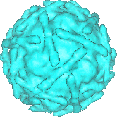 1057.gif
 http://ftp.pdbj.org/pub/emdb/structures/EMD-1057
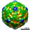


 Z (Sec.)
 Y (Row.)
 X (Col.)


 試料の構成要素
 Komagataella pastoris (菌類)
 解析
解析 電子顕微鏡法