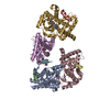
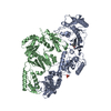
Journal: EBioMedicine / Year: 2020 Title: Structural insights and activating mutations in diverse pathologies define mechanisms of deregulation for phospholipase C gamma enzymes. Authors: Yang Liu / Tom D Bunney / Sakshi Khosa / Kévin Macé / Katharina Beckenbauer / Trevor Askwith / Sarah Maslen / Christopher Stubbs / Taiana M de Oliveira / Kasim Sader / Mark Skehel / Anne- ...Authors: Yang Liu / Tom D Bunney / Sakshi Khosa / Kévin Macé / Katharina Beckenbauer / Trevor Askwith / Sarah Maslen / Christopher Stubbs / Taiana M de Oliveira / Kasim Sader / Mark Skehel / Anne-Claude Gavin / Christopher Phillips / Matilda Katan / Abstract: BACKGROUND: PLCγ enzymes are key nodes in cellular signal transduction and their mutated and rare variants have been recently implicated in development of a range of diseases with unmet need ...BACKGROUND: PLCγ enzymes are key nodes in cellular signal transduction and their mutated and rare variants have been recently implicated in development of a range of diseases with unmet need including cancer, complex immune disorders, inflammation and neurodegenerative diseases. However, molecular nature of activation and the impact and dysregulation mechanisms by mutations, remain unclear; both are critically dependent on comprehensive characterization of the intact PLCγ enzymes. METHODS: For structural studies we applied cryo-EM, cross-linking mass spectrometry and hydrogen-deuterium exchange mass spectrometry. In parallel, we compiled mutations linked to main pathologies, ...METHODS: For structural studies we applied cryo-EM, cross-linking mass spectrometry and hydrogen-deuterium exchange mass spectrometry. In parallel, we compiled mutations linked to main pathologies, established their distribution and assessed their impact in cells and in vitro. FINDINGS: We define structure of a complex containing an intact, autoinhibited PLCγ1 and the intracellular part of FGFR1 and show that the interaction is centred on the nSH2 domain of PLCγ1. We ...FINDINGS: We define structure of a complex containing an intact, autoinhibited PLCγ1 and the intracellular part of FGFR1 and show that the interaction is centred on the nSH2 domain of PLCγ1. We define the architecture of PLCγ1 where an autoinhibitory interface involves the cSH2, spPH, TIM-barrel and C2 domains; this relative orientation occludes PLCγ1 access to its substrate. Based on this framework and functional characterization, the mechanism leading to an increase in PLCγ1 activity for the largest group of mutations is consistent with the major, direct impact on the autoinhibitory interface. INTERPRETATION: We reveal features of PLCγ enzymes that are important for determining their activation status. Targeting such features, as an alternative to targeting the PLC active site that has so ...INTERPRETATION: We reveal features of PLCγ enzymes that are important for determining their activation status. Targeting such features, as an alternative to targeting the PLC active site that has so far not been achieved for any PLC, could provide new routes for clinical interventions related to various pathologies driven by PLCγ deregulation. FUND: CR UK, MRC and AstaZeneca.
History
Deposition
Sep 4, 2019
-
Header (metadata) release
Jan 22, 2020
-
Map release
Jan 22, 2020
-
Update
Jan 29, 2020
-
Current status
Jan 29, 2020
Processing site: PDBe / Status: Released
-
Structure visualization
Movie
Surface view with section colored by density value
In the structure databanks used in Yorodumi, some data are registered as the other names, "COVID-19 virus" and "2019-nCoV". Here are the details of the virus and the list of structure data.
Jan 31, 2019. EMDB accession codes are about to change! (news from PDBe EMDB page)
EMDB accession codes are about to change! (news from PDBe EMDB page)
The allocation of 4 digits for EMDB accession codes will soon come to an end. Whilst these codes will remain in use, new EMDB accession codes will include an additional digit and will expand incrementally as the available range of codes is exhausted. The current 4-digit format prefixed with “EMD-” (i.e. EMD-XXXX) will advance to a 5-digit format (i.e. EMD-XXXXX), and so on. It is currently estimated that the 4-digit codes will be depleted around Spring 2019, at which point the 5-digit format will come into force.
The EM Navigator/Yorodumi systems omit the EMD- prefix.
Related info.:Q: What is EMD? / ID/Accession-code notation in Yorodumi/EM Navigator
Yorodumi is a browser for structure data from EMDB, PDB, SASBDB, etc.
This page is also the successor to EM Navigator detail page, and also detail information page/front-end page for Omokage search.
The word "yorodu" (or yorozu) is an old Japanese word meaning "ten thousand". "mi" (miru) is to see.
Related info.:EMDB / PDB / SASBDB / Comparison of 3 databanks / Yorodumi Search / Aug 31, 2016. New EM Navigator & Yorodumi / Yorodumi Papers / Jmol/JSmol / Function and homology information / Changes in new EM Navigator and Yorodumi
 Movie
Movie Controller

 Yorodumi
 Open data
 Basic information
 Map data
 Sample
 Homo sapiens (human)
 Authors
 United Kingdom, 1 items
  Citation

 Structure visualization
 Movie viewer
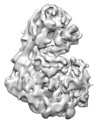
 Downloads & links
 emd_10288.png
 http://ftp.pdbj.org/pub/emdb/structures/EMD-10288
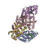


 Z (Sec.)
 Y (Row.)
 X (Col.)


 Sample components
 Mammalian 1 orthobornavirus
 Processing
 Electron microscopy
 FIELD EMISSION GUN
