 Movie
Movie Controller


+ Open data

- Basic information
| Entry | Database: EMDB / ID: EMD-10200 | |||||||||
|---|---|---|---|---|---|---|---|---|---|---|
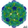
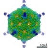
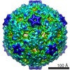
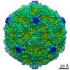
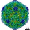
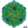
| Title | Structure of a marine algae virus of the order Picornavirales | |||||||||
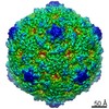
 Map data Map data | ||||||||||
 Sample Sample |
| |||||||||
 Keywords Keywords | Picornavirales / Marnaviridae / icosahedral virus / algae virus / jelly roll / VIRUS | |||||||||
| Function / homology | Capsid protein VP4, dicistrovirus / Cricket paralysis virus, VP4 / Dicistrovirus, capsid-polyprotein, C-terminal / CRPV capsid protein like / virion component / Picornavirus/Calicivirus coat protein / Viral coat protein subunit / Predicted structural protein / Predicted structural protein Function and homology information Function and homology information | |||||||||
| Biological species |  Chaetoceros tenuissimus RNA virus type-II Chaetoceros tenuissimus RNA virus type-II | |||||||||
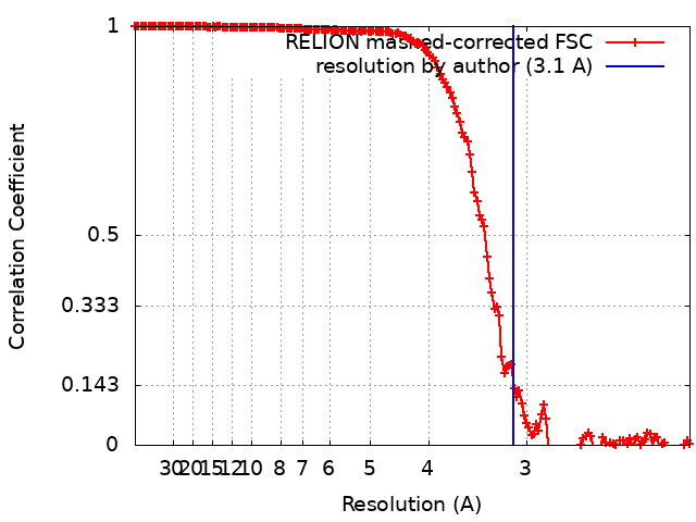
| Method | single particle reconstruction / cryo EM / Resolution: 3.1 Å | |||||||||
 Authors Authors | Munke A / Tomaru Y | |||||||||
| Funding support |  Sweden, 2 items Sweden, 2 items
| |||||||||
 Citation Citation | Journal: J Virol / Year: 2020 Title: Capsid Structure of a Marine Algal Virus of the Order . Authors: Anna Munke / Kei Kimura / Yuji Tomaru / Kenta Okamoto /  Abstract: The order includes viruses that infect different kinds of eukaryotes and that share similar properties. The capsid proteins (CPs) of viruses in the order that infect unicellular organisms, such as ...The order includes viruses that infect different kinds of eukaryotes and that share similar properties. The capsid proteins (CPs) of viruses in the order that infect unicellular organisms, such as algae, presumably possess certain characteristics that have changed little over the course of evolution, and thus these viruses may resemble the ancestor in some respects. Herein, we present the capsid structure of RNA virus type II (CtenRNAV-II) determined using cryo-electron microscopy at a resolution of 3.1 Å, the first alga virus belonging to the family of the order A structural comparison to related invertebrate and vertebrate viruses revealed a unique surface loop of the major CP VP1 that had not been observed previously, and further, revealed that another VP1 loop obscures the so-called canyon, which is a host-receptor binding site for many of the mammalian viruses. VP2 has an N-terminal tail, which has previously been reported as a primordial feature of viruses. The above-mentioned and other critical structural features provide new insights on three long-standing theories about : (i) the canyon hypothesis, (ii) the primordial VP2 domain swap, and (iii) the hypothesis that alga viruses could share characteristics with the ancestor. Identifying the acquired structural traits in virus capsids is important for elucidating what functions are essential among viruses that infect different hosts. The viruses infect a broad spectrum of hosts, ranging from unicellular algae to insects and mammals and include many human pathogens. Those viruses that infect unicellular protists, such as algae, are likely to have undergone fewer structural changes during the course of evolution compared to those viruses that infect multicellular eukaryotes and thus still share some characteristics with the ancestor. This article describes the first atomic capsid structure of an alga , CtenRNAV-II. A comparison to capsid structures of the related invertebrate and vertebrate viruses identified a number of structural traits that have been functionally acquired or lost during the course of evolution. These observations provide new insights on past theories on the viability and evolution of viruses. | |||||||||
| History |
|
- Structure visualization
| Movie |
Movie viewer |
|---|---|
| Structure viewer | EM map: SurfViewMolmilJmol/JSmol |
| Supplemental images |

- Downloads & links
-EMDB archive
| Map data | emd_10200.map.gz | 63.2 MB | EMDB map data format | |
|---|---|---|---|---|
| Header (meta data) | emd-10200-v30.xmlemd-10200.xml | 15.1 KB 15.1 KB | Display Display | EMDB header |
| FSC (resolution estimation) | emd_10200_fsc.xml | 15.9 KB | Display | FSC data file |
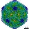
| Images | 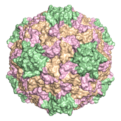 emd_10200.png emd_10200.png | 234.2 KB | ||
| Filedesc metadata | emd-10200.cif.gz | 5.8 KB | ||
| Archive directory |  http://ftp.pdbj.org/pub/emdb/structures/EMD-10200ftp://ftp.pdbj.org/pub/emdb/structures/EMD-10200 http://ftp.pdbj.org/pub/emdb/structures/EMD-10200ftp://ftp.pdbj.org/pub/emdb/structures/EMD-10200 | HTTPS FTP |
-Related structure data
| Related structure data |  6shlMC M: atomic model generated by this map C: citing same article ( |
|---|---|
| Similar structure data |

-Links
| EMDB pages | EMDB (EBI/PDBe) / EMDataResource |
|---|
-Map
| File | Download / File: emd_10200.map.gz / Format: CCP4 / Size: 347.6 MB / Type: IMAGE STORED AS FLOATING POINT NUMBER (4 BYTES) | ||||||||||||||||||||||||||||||||||||||||||||||||||||||||||||
|---|---|---|---|---|---|---|---|---|---|---|---|---|---|---|---|---|---|---|---|---|---|---|---|---|---|---|---|---|---|---|---|---|---|---|---|---|---|---|---|---|---|---|---|---|---|---|---|---|---|---|---|---|---|---|---|---|---|---|---|---|---|
| Projections & slices | Image control
Images are generated by Spider. | ||||||||||||||||||||||||||||||||||||||||||||||||||||||||||||
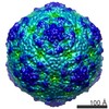
| Voxel size | X=Y=Z: 1.06 Å | ||||||||||||||||||||||||||||||||||||||||||||||||||||||||||||
| Density |
| ||||||||||||||||||||||||||||||||||||||||||||||||||||||||||||
| Symmetry | Space group: 1 | ||||||||||||||||||||||||||||||||||||||||||||||||||||||||||||
| Details | EMDB XML:
CCP4 map header:
| ||||||||||||||||||||||||||||||||||||||||||||||||||||||||||||
 Z (Sec.)
 Y (Row.)
 X (Col.)


-Supplemental data
- Sample components
-Entire : Chaetoceros tenuissimus RNA virus type-II
| Entire | Name: Chaetoceros tenuissimus RNA virus type-II |
|---|---|
| Components |
|
-Supramolecule #1: Chaetoceros tenuissimus RNA virus type-II
| Supramolecule | Name: Chaetoceros tenuissimus RNA virus type-II / type: virus / ID: 1 / Parent: 0 / Macromolecule list: all / NCBI-ID: 1516128 / Sci species name: Chaetoceros tenuissimus RNA virus type-II / Virus type: VIRION / Virus isolate: STRAIN / Virus enveloped: No / Virus empty: No |
|---|---|
| Host (natural) | Organism:  Chaetoceros tenuissimus (Diatom) Chaetoceros tenuissimus (Diatom) |
| Virus shell | Shell ID: 1 / Name: Capsid / T number (triangulation number): 3 |
-Macromolecule #1: VP1
| Macromolecule | Name: VP1 / type: protein_or_peptide / ID: 1 / Number of copies: 1 / Enantiomer: LEVO |
|---|---|
| Source (natural) | Organism: Chaetoceros tenuissimus RNA virus type-II |
| Molecular weight | Theoretical: 29.740906 KDa |
| Sequence | String: DNSNNPVGGN PIDNYGTEHA PLLKEDNQYL VYQGERIVSF KDLLRRYQYL NSYWPQETGS GFRYYTLDSP GMPIYRGWDP NGIDQGQDS TAGNSPYNFC SMTLLNYLAP AFVCQRGSLR HKWVTAGARV NSTASVLSAT RHGVLFPLPL AETAHPLDNA L VGDRRSEL ...String: DNSNNPVGGN PIDNYGTEHA PLLKEDNQYL VYQGERIVSF KDLLRRYQYL NSYWPQETGS GFRYYTLDSP GMPIYRGWDP NGIDQGQDS TAGNSPYNFC SMTLLNYLAP AFVCQRGSLR HKWVTAGARV NSTASVLSAT RHGVLFPLPL AETAHPLDNA L VGDRRSEL QEMQRSRLNG TAITPVRLNN TLEIELPYYS IGQRFHASRF LDLAGTGDTQ GVEIACEISD GGNDANYRLD QF VSVGEDF TLGMFVGAPI MYFYNDPTAT UniProtKB: Predicted structural protein |
-Macromolecule #2: VP2
| Macromolecule | Name: VP2 / type: protein_or_peptide / ID: 2 / Number of copies: 1 / Enantiomer: LEVO |
|---|---|
| Source (natural) | Organism: Chaetoceros tenuissimus RNA virus type-II |
| Molecular weight | Theoretical: 25.911801 KDa |
| Sequence | String: PSANDGPSFT TSKTSQNTTS ENVHFVDGDT PWTYDVAATP DETSKLSGFD DAGLGEFLSR PIKIQQYQWT PGVQLFQTFN PWSDYFGNA DVLEKINRFR NLRCKLCLKV LINGNSFYYG RALLSYNPYL RNDQVTVNRS FFIQDLIAAS NKPHILLDPC S SEGGQMCL ...String: PSANDGPSFT TSKTSQNTTS ENVHFVDGDT PWTYDVAATP DETSKLSGFD DAGLGEFLSR PIKIQQYQWT PGVQLFQTFN PWSDYFGNA DVLEKINRFR NLRCKLCLKV LINGNSFYYG RALLSYNPYL RNDQVTVNRS FFIQDLIAAS NKPHILLDPC S SEGGQMCL PFIWPENYLD ITSTGWEDQM GECIIHDFDV LRHANGGTDP ITVSIFAWAE DVSLLIPTTV AAQ UniProtKB: Predicted structural protein |
-Macromolecule #3: VP3
| Macromolecule | Name: VP3 / type: protein_or_peptide / ID: 3 / Number of copies: 1 / Enantiomer: LEVO |
|---|---|
| Source (natural) | Organism: Chaetoceros tenuissimus RNA virus type-II |
| Molecular weight | Theoretical: 28.508686 KDa |
| Sequence | String: SRPAVLSDIQ PYVPRYCGNL ANSDAPETVN KLSVDSKNEL TIDTRTMGLG GADELTIHSI ASRMTFWRQF DWPESAVTDT LLASMSVQP FCIDTVTASP VTEIHSTALA FASAPFETWQ GSIKFHFKVV CSEYHRGRLR LVYNPLTNNA GPVAFNQVYS T TIDISNDR ...String: SRPAVLSDIQ PYVPRYCGNL ANSDAPETVN KLSVDSKNEL TIDTRTMGLG GADELTIHSI ASRMTFWRQF DWPESAVTDT LLASMSVQP FCIDTVTASP VTEIHSTALA FASAPFETWQ GSIKFHFKVV CSEYHRGRLR LVYNPLTNNA GPVAFNQVYS T TIDISNDR EFDYECKWTD IRAWNACIGI DGATSATFFN TAAAVTGGTP FDNGTLSVYV VNELATPSTA AADVKVQVWV SA GDDFAVA VPGVGLSQLS YFQQQ UniProtKB: Predicted structural protein |
-Macromolecule #4: VP4
| Macromolecule | Name: VP4 / type: protein_or_peptide / ID: 4 / Number of copies: 1 / Enantiomer: LEVO |
|---|---|
| Source (natural) | Organism: Chaetoceros tenuissimus RNA virus type-II |
| Molecular weight | Theoretical: 5.406366 KDa |
| Sequence | String: EFKHDGLISK PASAVAKAAD ALSMIPYIAP YAKATSMVAD KIGKIARIFG Y UniProtKB: Predicted structural protein |
-Experimental details
-Structure determination
| Method | cryo EM |
|---|---|
 Processing Processing | single particle reconstruction |
| Aggregation state | particle |
-Sample preparation
| Buffer | pH: 7.4 |
|---|---|
| Grid | Model: Quantifoil R2/2 / Material: COPPER / Mesh: 300 / Support film - Material: CARBON / Support film - topology: HOLEY / Pretreatment - Type: GLOW DISCHARGE |
| Vitrification | Cryogen name: ETHANE / Chamber humidity: 100 % / Chamber temperature: 277 K / Instrument: FEI VITROBOT MARK IV |
- Electron microscopy
| Microscope | FEI TITAN KRIOS |
|---|---|
| Image recording | Film or detector model: GATAN K2 SUMMIT (4k x 4k) / Detector mode: COUNTING / Number grids imaged: 1 / Number real images: 2592 / Average exposure time: 8.0 sec. / Average electron dose: 29.0 e/Å2 |
| Electron beam | Acceleration voltage: 300 kV / Electron source:  FIELD EMISSION GUN FIELD EMISSION GUN |
| Electron optics | Illumination mode: FLOOD BEAM / Imaging mode: BRIGHT FIELD / Nominal defocus max: 3.0 µm / Nominal defocus min: 1.0 µm / Nominal magnification: 140000 |
| Sample stage | Specimen holder model: FEI TITAN KRIOS AUTOGRID HOLDER / Cooling holder cryogen: NITROGEN |
| Experimental equipment |  Model: Titan Krios / Image courtesy: FEI Company |
+Image processing
-Atomic model buiding 1
| Refinement | Space: REAL / Protocol: BACKBONE TRACE |
|---|---|
| Output model | PDB-6shl: |
