 ムービー
ムービー コントローラー


+ データを開く

- 基本情報
基本情報
| 登録情報 | データベース: EMDB / ID: EMD-1020 | |||||||||
|---|---|---|---|---|---|---|---|---|---|---|
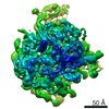
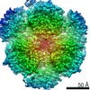
| タイトル | Structure of a viral DNA gatekeeper at 10 A resolution by cryo-electron microscopy. | |||||||||
 マップデータ マップデータ | Viral DNA gatekeeper. Centre of symmetry : 50.0, 50.0, 50.0 | |||||||||
 試料 試料 |
| |||||||||
| 生物種 |  Bacillus phage SPP1 (ファージ) Bacillus phage SPP1 (ファージ) | |||||||||
| 手法 | 単粒子再構成法 / クライオ電子顕微鏡法 / 解像度: 10.0 Å | |||||||||
 データ登録者 データ登録者 | Orlova EV / Gowen B / Droege A / Stiege A / Weise F / Lurz R / van Heel M / Tavares P | |||||||||
 引用 引用 | ジャーナル: EMBO J / 年: 2003 タイトル: Structure of a viral DNA gatekeeper at 10 A resolution by cryo-electron microscopy. 著者: Elena V Orlova / Brent Gowen / Anja Dröge / Asita Stiege / Frank Weise / Rudi Lurz / Marin van Heel / Paulo Tavares /  要旨: In tailed bacteriophages and herpes viruses, the viral DNA is packaged through the portal protein channel. Channel closure is essential to prevent DNA release after packaging. Here we present the ...In tailed bacteriophages and herpes viruses, the viral DNA is packaged through the portal protein channel. Channel closure is essential to prevent DNA release after packaging. Here we present the connector structure from bacteriophage SPP1 using cryo-electron microscopy and single particle analysis. The multiprotein complex comprises the portal protein gp6 and the head completion proteins gp15 and gp16. Although we show that gp6 in the connector has a fold similar to that of the isolated portal protein, we observe conformational changes in the region of gp6 exposed to the DNA-packaging ATPase and to gp15. This reorganization does not cause closure of the channel. The connector channel traverses the full height of gp6 and gp15, but it is closed by gp16 at the bottom of the complex. Gp16 acts as a valve whose closure prevents DNA leakage, while its opening is required for DNA release upon interaction of the virus with its host. | |||||||||
| 履歴 |
|
- 構造の表示
構造の表示
| ムービー |
 ムービービューア ムービービューア |
|---|---|
| 構造ビューア | EMマップ: SurfViewMolmilJmol/JSmol |
| 添付画像 |


- ダウンロードとリンク
-EMDBアーカイブ
| マップデータ | emd_1020.map.gz | 1.3 MB | EMDBマップデータ形式 | |
|---|---|---|---|---|
| ヘッダ (付随情報) | emd-1020-v30.xmlemd-1020.xml | 9.5 KB 9.5 KB | 表示 表示 | EMDBヘッダ |
| 画像 | 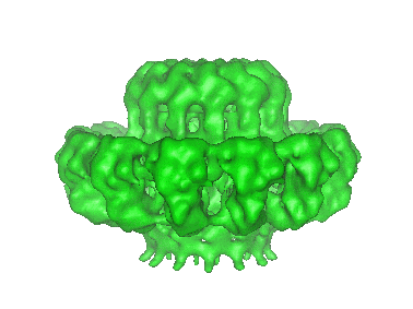 1020.gifemd_1020.tifemd_emd_1020.tif 1020.gifemd_1020.tifemd_emd_1020.tif | 23.5 KB 588.8 KB 588.8 KB | ||
| その他 | Resolution_Portal_Connector.tif | 143.8 KB | ||
| アーカイブディレクトリ |  http://ftp.pdbj.org/pub/emdb/structures/EMD-1020ftp://ftp.pdbj.org/pub/emdb/structures/EMD-1020 http://ftp.pdbj.org/pub/emdb/structures/EMD-1020ftp://ftp.pdbj.org/pub/emdb/structures/EMD-1020 | HTTPS FTP |
-検証レポート
| 文書・要旨 | emd_1020_validation.pdf.gz | 235 KB | 表示 | EMDB検証レポート |
|---|---|---|---|---|
| 文書・詳細版 | emd_1020_full_validation.pdf.gz | 234.1 KB | 表示 | |
| XML形式データ | emd_1020_validation.xml.gz | 5.3 KB | 表示 | |
| アーカイブディレクトリ | https://ftp.pdbj.org/pub/emdb/validation_reports/EMD-1020ftp://ftp.pdbj.org/pub/emdb/validation_reports/EMD-1020 | HTTPS FTP |
-関連構造データ
-リンク
| EMDBのページ | EMDB (EBI/PDBe) / EMDataResource |
|---|
-マップ
| ファイル | ダウンロード / ファイル: emd_1020.map.gz / 形式: CCP4 / 大きさ: 3.7 MB / タイプ: IMAGE STORED AS FLOATING POINT NUMBER (4 BYTES) | ||||||||||||||||||||||||||||||||||||||||||||||||||||||||||||
|---|---|---|---|---|---|---|---|---|---|---|---|---|---|---|---|---|---|---|---|---|---|---|---|---|---|---|---|---|---|---|---|---|---|---|---|---|---|---|---|---|---|---|---|---|---|---|---|---|---|---|---|---|---|---|---|---|---|---|---|---|---|
| 注釈 | Viral DNA gatekeeper. Centre of symmetry : 50.0, 50.0, 50.0 | ||||||||||||||||||||||||||||||||||||||||||||||||||||||||||||
| 投影像・断面図 | 画像のコントロール
画像は Spider により作成 | ||||||||||||||||||||||||||||||||||||||||||||||||||||||||||||
| ボクセルのサイズ | X=Y=Z: 2.1 Å | ||||||||||||||||||||||||||||||||||||||||||||||||||||||||||||
| 密度 |
| ||||||||||||||||||||||||||||||||||||||||||||||||||||||||||||
| 対称性 | 空間群: 1 | ||||||||||||||||||||||||||||||||||||||||||||||||||||||||||||
| 詳細 | EMDB XML:
CCP4マップ ヘッダ情報:
| ||||||||||||||||||||||||||||||||||||||||||||||||||||||||||||
 Z (Sec.)
 Y (Row.)
 X (Col.)


-添付データ
- 試料の構成要素
-全体 : Bacteriophage SPP1 portal protein gp6
| 全体 | 名称: Bacteriophage SPP1 portal protein gp6 |
|---|---|
| 要素 |
|
-超分子 #1000: Bacteriophage SPP1 portal protein gp6
| 超分子 | 名称: Bacteriophage SPP1 portal protein gp6 / タイプ: sample / ID: 1000 集合状態: isolated portal protein has 13- fold cyclical symmetry Number unique components: 1 |
|---|---|
| 分子量 | 実験値: 57.3 KDa / 理論値: 57.3 KDa |
-分子 #1: Bacteriophage SPP1 portal protein gp6
| 分子 | 名称: Bacteriophage SPP1 portal protein gp6 / タイプ: protein_or_peptide / ID: 1 / Name.synonym: naive gp6 / コピー数: 13 / 集合状態: C13 / 組換発現: Yes |
|---|---|
| 由来(天然) | 生物種: Bacillus phage SPP1 (ファージ) / 別称: Bacteriophage SPP1 |
| 組換発現 | 生物種:  |
-実験情報
-構造解析
| 手法 | クライオ電子顕微鏡法 |
|---|---|
 解析 解析 | 単粒子再構成法 |
| 試料の集合状態 | particle |
-試料調製
| 濃度 | 1.5 mg/mL |
|---|---|
| 緩衝液 | pH: 7.5 |
| 凍結 | 凍結剤: ETHANE / 装置: HOMEMADE PLUNGER / 詳細: Vitrification instrument: Standard plunger / 手法: Blot about 2-3 seconds before plunging. |
- 電子顕微鏡法
| 顕微鏡 | FEI/PHILIPS CM200FEG/ST |
|---|---|
| 日付 | 1999年1月1日 |
| 撮影 | カテゴリ: FILM / フィルム・検出器のモデル: KODAK SO-163 FILM / デジタル化 - スキャナー: PATCHWORK DENSITOMETER / デジタル化 - サンプリング間隔: 10.2 µm / 実像数: 15 / 平均電子線量: 10 e/Å2 詳細: Images were digitised using a patch work densitometer. Od range: 1.6 / ビット/ピクセル: 8 |
| 電子線 | 加速電圧: 200 kV / 電子線源:  FIELD EMISSION GUN FIELD EMISSION GUN |
| 電子光学系 | 倍率(補正後): 48600 / 照射モード: FLOOD BEAM / 撮影モード: BRIGHT FIELD / Cs: 2.1 mm / 最大 デフォーカス(公称値): 3.0 µm / 最小 デフォーカス(公称値): 1.7 µm / 倍率(公称値): 50000 |
| 試料ステージ | 試料ホルダー: side-entry / 試料ホルダーモデル: GATAN LIQUID NITROGEN |
-画像解析
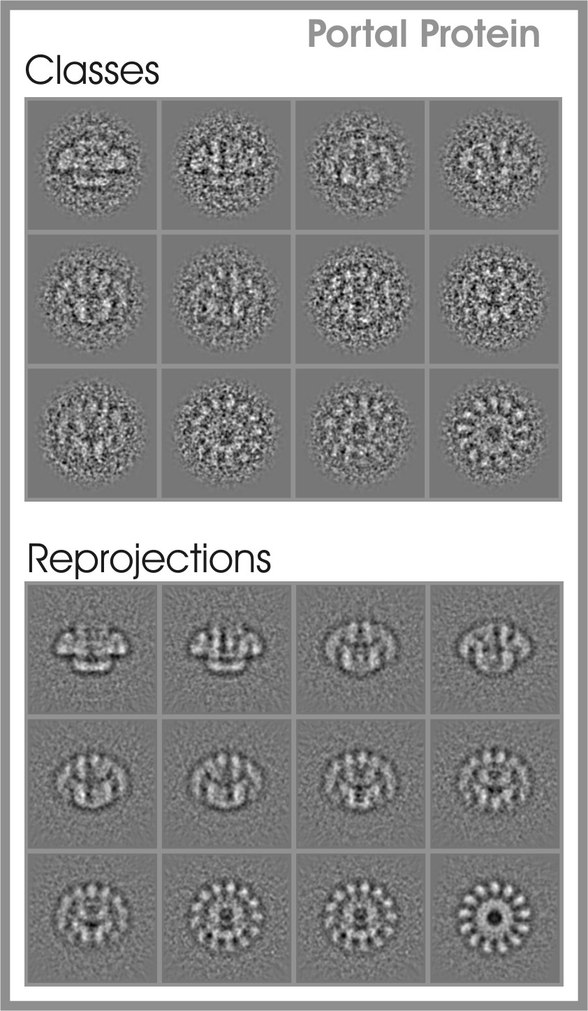
| 詳細 | The number of classes analysed was 650. In the final reconstruction were used only 430. |
|---|---|
| CTF補正 | 詳細: CTF correction of each particle |
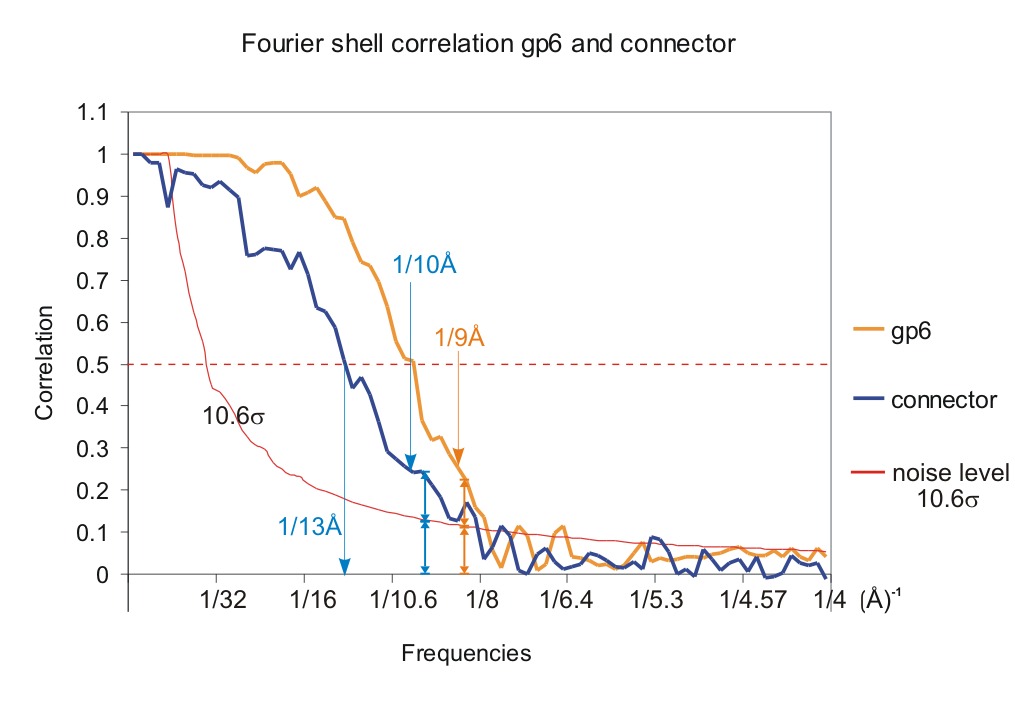
| 最終 再構成 | 想定した対称性 - 点群: C13 (13回回転対称) / アルゴリズム: OTHER / 解像度のタイプ: BY AUTHOR / 解像度: 10.0 Å / 解像度の算出法: OTHER / ソフトウェア - 名称: IMAGIC / 使用した粒子像数: 8000 |
| 最終 2次元分類 | クラス数: 430 |
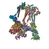
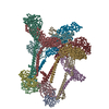
-原子モデル構築 1
| ソフトウェア | 名称: Molrep |
|---|---|
| 詳細 | Protocol: Angular reconstitution |
