Movie
Movie Controller

[English] 日本語
 Yorodumi
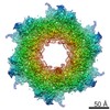
- EMDB-10040: Inner membrane ring and secretin N0 N1 domains of the Shigella ty... -
+ Open data

- Basic information
| Entry | Database: EMDB / ID: EMD-10040 | |||||||||
|---|---|---|---|---|---|---|---|---|---|---|
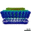
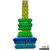
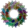
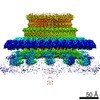
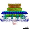
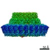
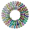
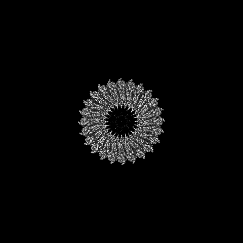
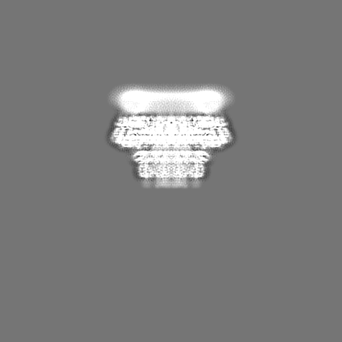
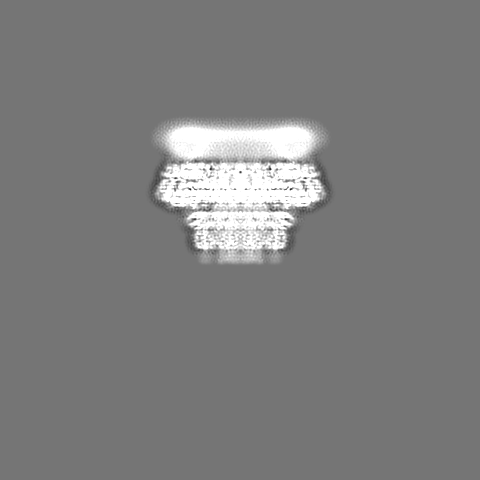
| Title | Inner membrane ring and secretin N0 N1 domains of the Shigella type 3 secretion system | |||||||||
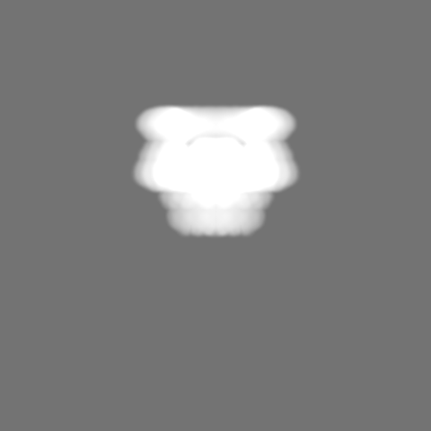
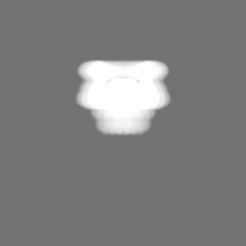
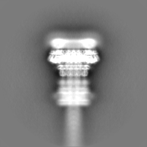
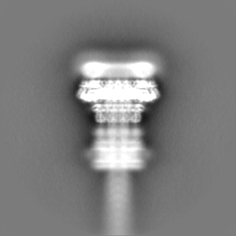
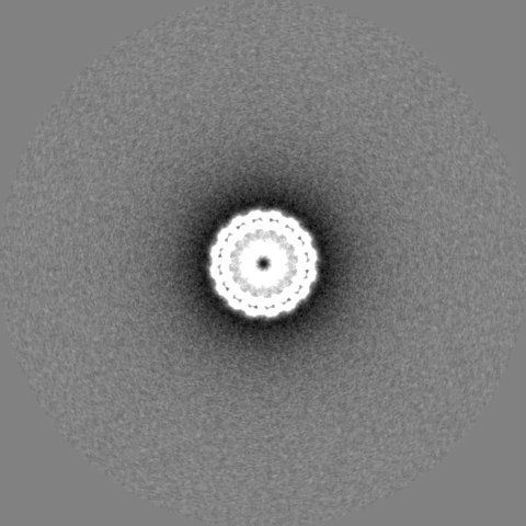
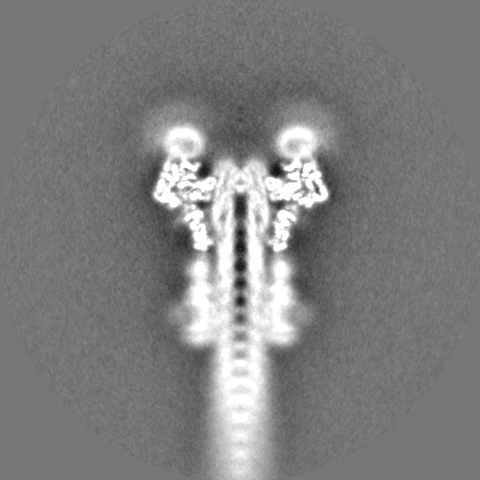
 Map data Map data | Masked C8 map of IM ring and connector, post-processed filtered at 3.86 A and sharpened (b=-120) | |||||||||
 Sample Sample |
| |||||||||
 Keywords Keywords | type 3 secretion system / shigella / secretin / beta-sheet augmentation / PROTEIN TRANSPORT | |||||||||
| Function / homology |  Function and homology information Function and homology informationtype III protein secretion system complex / type II protein secretion system complex / protein secretion by the type III secretion system / cell outer membrane / plasma membrane Similarity search - Function | |||||||||
| Biological species |  Shigella flexneri (bacteria) Shigella flexneri (bacteria) | |||||||||
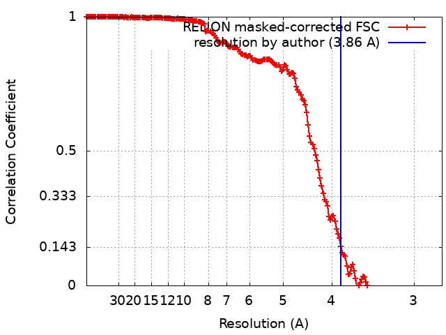
| Method | single particle reconstruction / cryo EM / Resolution: 3.86 Å | |||||||||
 Authors Authors | Lunelli M / Kamprad A | |||||||||
| Funding support | 2 items
| |||||||||
 Citation Citation | Journal: PLoS Pathog / Year: 2020 Title: Cryo-EM structure of the Shigella type III needle complex. Authors: Michele Lunelli / Antje Kamprad / Jörg Bürger / Thorsten Mielke / Christian M T Spahn / Michael Kolbe /  Abstract: The Type III Secretion Systems (T3SS) needle complex is a conserved syringe-shaped protein translocation nanomachine with a mass of about 3.5 MDa essential for the survival and virulence of many Gram- ...The Type III Secretion Systems (T3SS) needle complex is a conserved syringe-shaped protein translocation nanomachine with a mass of about 3.5 MDa essential for the survival and virulence of many Gram-negative bacterial pathogens. This system is composed of a membrane-embedded basal body and an extracellular needle that deliver effector proteins into host cells. High-resolution structures of the T3SS from different organisms and infection stages are needed to understand the underlying molecular mechanisms of effector translocation. Here, we present the cryo-electron microscopy structure of the isolated Shigella T3SS needle complex. The inner membrane (IM) region of the basal body adopts 24-fold rotational symmetry and forms a channel system that connects the bacterial periplasm with the export apparatus cage. The secretin oligomer adopts a heterogeneous architecture with 16- and 15-fold cyclic symmetry in the periplasmic N-terminal connector and C-terminal outer membrane ring, respectively. Two out of three IM subunits bind the secretin connector via a β-sheet augmentation. The cryo-EM map also reveals the helical architecture of the export apparatus core, the inner rod, the needle and their intervening interfaces. | |||||||||
| History |
|
- Structure visualization
| Movie |
Movie viewer |
|---|---|
| Structure viewer | EM map: SurfViewMolmilJmol/JSmol |
| Supplemental images |

- Downloads & links
-EMDB archive
| Map data | emd_10040.map.gz | 30.1 MB | EMDB map data format | |
|---|---|---|---|---|
| Header (meta data) | emd-10040-v30.xmlemd-10040.xml | 22.8 KB 22.8 KB | Display Display | EMDB header |
| FSC (resolution estimation) | emd_10040_fsc.xml | 17 KB | Display | FSC data file |
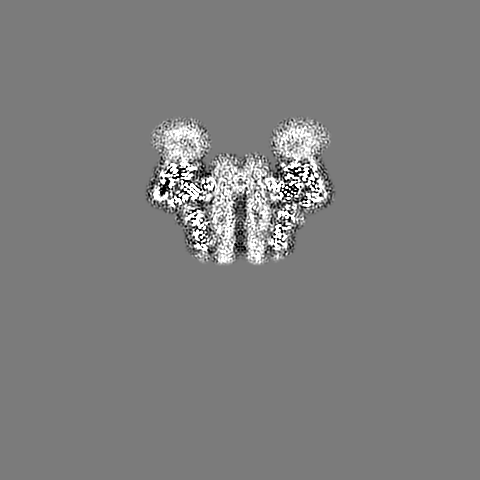
| Images | 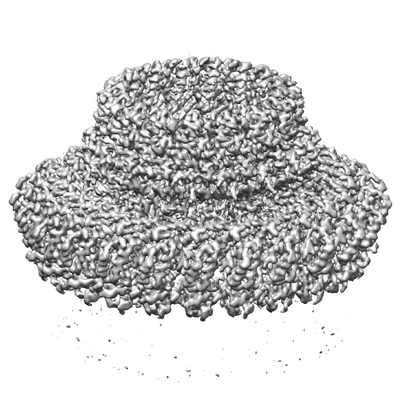 emd_10040.png emd_10040.png | 73.6 KB | ||
| Masks | emd_10040_msk_1.map | 421.9 MB | Mask map | |
| Filedesc metadata | emd-10040.cif.gz | 6.6 KB | ||
| Others | emd_10040_additional.map.gzemd_10040_half_map_1.map.gzemd_10040_half_map_2.map.gz | 30.7 MB 330.4 MB 330.5 MB | ||
| Archive directory |  http://ftp.pdbj.org/pub/emdb/structures/EMD-10040ftp://ftp.pdbj.org/pub/emdb/structures/EMD-10040 http://ftp.pdbj.org/pub/emdb/structures/EMD-10040ftp://ftp.pdbj.org/pub/emdb/structures/EMD-10040 | HTTPS FTP |
-Validation report
| Summary document | emd_10040_validation.pdf.gz | 368 KB | Display | EMDB validaton report |
|---|---|---|---|---|
| Full document | emd_10040_full_validation.pdf.gz | 367.1 KB | Display | |
| Data in XML | emd_10040_validation.xml.gz | 22 KB | Display | |
| Arichive directory | https://ftp.pdbj.org/pub/emdb/validation_reports/EMD-10040ftp://ftp.pdbj.org/pub/emdb/validation_reports/EMD-10040 | HTTPS FTP |
-Related structure data
| Related structure data | 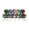 6rwkMC 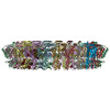 6rwxC 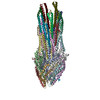 6rwyC C: citing same article ( M: atomic model generated by this map |
|---|---|
| Similar structure data |
-Links
| EMDB pages | EMDB (EBI/PDBe) / EMDataResource |
|---|---|
| Related items in Molecule of the Month |

-Map
| File | Download / File: emd_10040.map.gz / Format: CCP4 / Size: 421.9 MB / Type: IMAGE STORED AS FLOATING POINT NUMBER (4 BYTES) | ||||||||||||||||||||||||||||||||||||||||||||||||||||||||||||
|---|---|---|---|---|---|---|---|---|---|---|---|---|---|---|---|---|---|---|---|---|---|---|---|---|---|---|---|---|---|---|---|---|---|---|---|---|---|---|---|---|---|---|---|---|---|---|---|---|---|---|---|---|---|---|---|---|---|---|---|---|---|
| Annotation | Masked C8 map of IM ring and connector, post-processed filtered at 3.86 A and sharpened (b=-120) | ||||||||||||||||||||||||||||||||||||||||||||||||||||||||||||
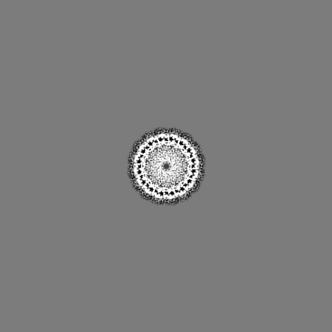
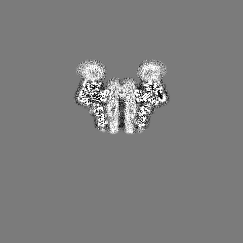
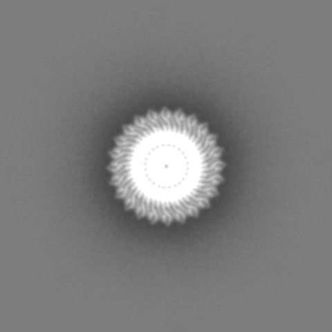
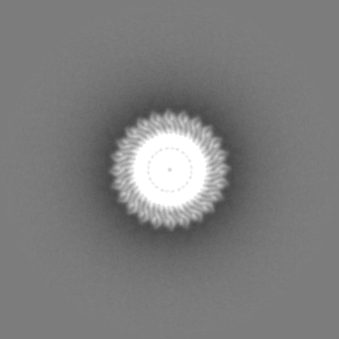
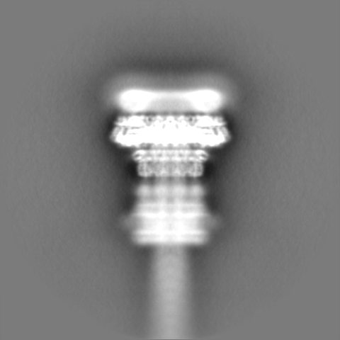
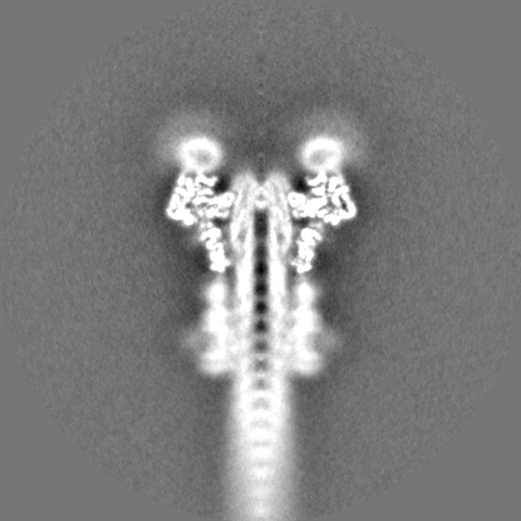
| Projections & slices | Image control
Images are generated by Spider. | ||||||||||||||||||||||||||||||||||||||||||||||||||||||||||||
| Voxel size | X=Y=Z: 1.38369 Å | ||||||||||||||||||||||||||||||||||||||||||||||||||||||||||||
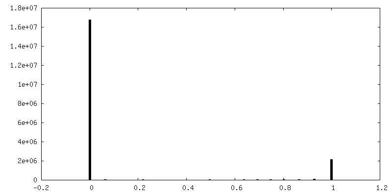
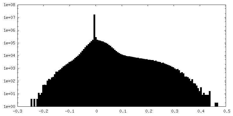
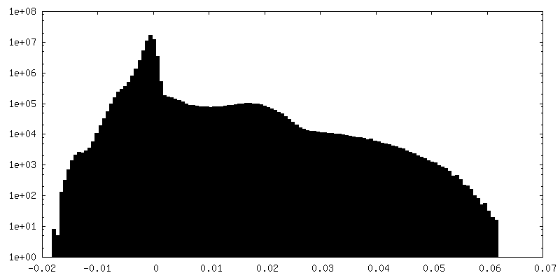
| Density |
| ||||||||||||||||||||||||||||||||||||||||||||||||||||||||||||
| Symmetry | Space group: 1 | ||||||||||||||||||||||||||||||||||||||||||||||||||||||||||||
| Details | EMDB XML:
CCP4 map header:
| ||||||||||||||||||||||||||||||||||||||||||||||||||||||||||||
 Z (Sec.)
 Y (Row.)
 X (Col.)


-Supplemental data
-Mask #1
| File | emd_10040_msk_1.map | ||||||||||||
|---|---|---|---|---|---|---|---|---|---|---|---|---|---|
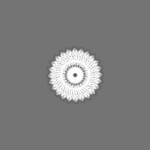
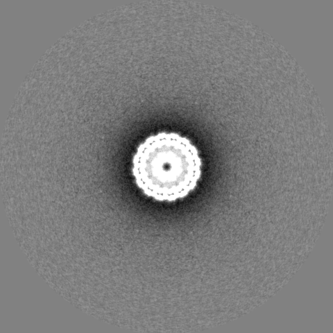
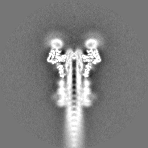
| Projections & Slices |
| ||||||||||||
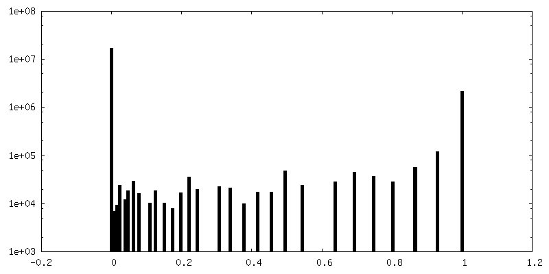
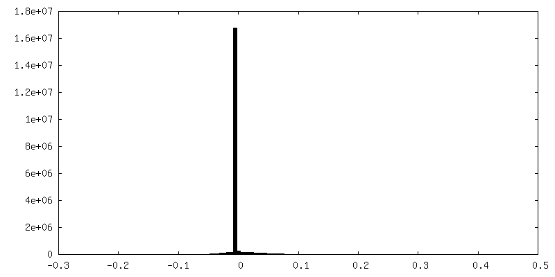
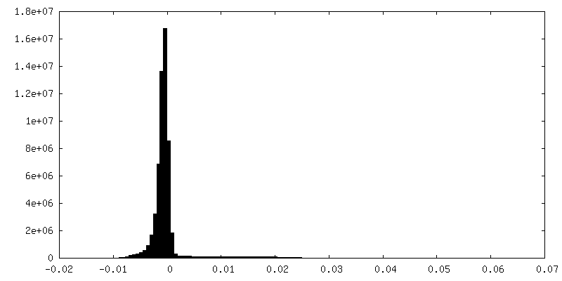
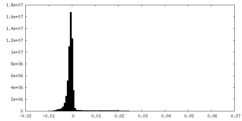
| Density Histograms |


-Additional map: Masked C8 map of IM ring and connector,...
| File | emd_10040_additional.map | ||||||||||||
|---|---|---|---|---|---|---|---|---|---|---|---|---|---|
| Annotation | Masked C8 map of IM ring and connector, post-processed filtered at 3.5 A and sharpened (b=-120), used for model building and refinement. | ||||||||||||
| Projections & Slices |
| ||||||||||||
| Density Histograms |
-Half map: Unfiltered half map of C8 reconstruction of IM ring and connector.
| File | emd_10040_half_map_1.map | ||||||||||||
|---|---|---|---|---|---|---|---|---|---|---|---|---|---|
| Annotation | Unfiltered half map of C8 reconstruction of IM ring and connector. | ||||||||||||
| Projections & Slices |
| ||||||||||||
| Density Histograms |


-Half map: Unfiltered half map of C8 reconstruction of IM ring and connector.
| File | emd_10040_half_map_2.map | ||||||||||||
|---|---|---|---|---|---|---|---|---|---|---|---|---|---|
| Annotation | Unfiltered half map of C8 reconstruction of IM ring and connector. | ||||||||||||
| Projections & Slices |
| ||||||||||||
| Density Histograms |

- Sample components
-Entire : Oligomer of the secretin N0 and N1 domains in complex with the in...
| Entire | Name: Oligomer of the secretin N0 and N1 domains in complex with the inner membrane ring of the Shigella type 3 secretion system |
|---|---|
| Components |
|
-Supramolecule #1: Oligomer of the secretin N0 and N1 domains in complex with the in...
| Supramolecule | Name: Oligomer of the secretin N0 and N1 domains in complex with the inner membrane ring of the Shigella type 3 secretion system type: complex / ID: 1 / Parent: 0 / Macromolecule list: all Details: Focused reconstruction from isolated needle complex of the Shigella type 3 secretion system |
|---|---|
| Source (natural) | Organism: Shigella flexneri (bacteria) / Strain: M90T / Location in cell: Membrane |
-Macromolecule #1: Outer membrane protein MxiD
| Macromolecule | Name: Outer membrane protein MxiD / type: protein_or_peptide / ID: 1 Details: MxiD N0 and N1 N-terminal domains in complex with MxiG C-terminal domain Number of copies: 16 / Enantiomer: LEVO |
|---|---|
| Source (natural) | Organism: Shigella flexneri (bacteria) |
| Molecular weight | Theoretical: 63.230414 KDa |
| Sequence | String: MKKFNIKSLT LLIVLLPLIV NANNIDSHLL EQNDIAKYVA QSDTVGSFFE RFSALLNYPI VVSKQAAKKR ISGEFDLSNP EEMLEKLTL LVGLIWYKDG NALYIYDSGE LISKVILLEN ISLNYLIQYL KDANLYDHRY PIRGNISDKT FYISGPPALV E LVANTATL ...String: MKKFNIKSLT LLIVLLPLIV NANNIDSHLL EQNDIAKYVA QSDTVGSFFE RFSALLNYPI VVSKQAAKKR ISGEFDLSNP EEMLEKLTL LVGLIWYKDG NALYIYDSGE LISKVILLEN ISLNYLIQYL KDANLYDHRY PIRGNISDKT FYISGPPALV E LVANTATL LDKQVSSIGT DKVNFGVIKL KNTFVSDRTY NMRGEDIVIP GVATVVERLL NNGKALSNRQ AQNDPMPPFN IT QKVSEDS NDFSFSSVTN SSILEDVSLI AYPETNSILV KGNDQQIQII RDIITQLDVA KRHIELSLWI IDIDKSELNN LGV NWQGTA SFGDSFGASF NMSSSASIST LDGNKFIASV MALNQKKKAN VVSRPVILTQ ENIPAIFDNN RTFYVSLVGE RNSS LEHVT YGTLINVIPR FSSRGQIEMS LTIEDGTGNS QSNYNYNNEN TSVLPEVGRT KISTIARVPQ GKSLLIGGYT HETNS NEII SIPFLSSIPV IGNVFKYKTS NISNIVRVFL IQPREIKESS YYNTAEYKSL ISEREIQKTT QIIPSETTLL EDEKSL VSY LNY UniProtKB: Type 3 secretion system secretin |
-Macromolecule #2: Protein MxiG
| Macromolecule | Name: Protein MxiG / type: protein_or_peptide / ID: 2 Details: MxiG C-terminal domain in complex with secretin MxiD Number of copies: 16 / Enantiomer: LEVO |
|---|---|
| Source (natural) | Organism: Shigella flexneri (bacteria) |
| Molecular weight | Theoretical: 43.053844 KDa |
| Sequence | String: MSEAKNSNLA PFRLLVKLTN GVGDEFPLYY GNNLIVLGRT IETLEFGNDN FPENIIPVTD SKSDGIIYLT ISKDNICQFS DEKGEQIDI NSQFNSFEYD GISFHLKNMR EDKSRGHILN GMYKNHSVFF FFAVIVVLII IFSLSLKKDE VKEIAEIIDD K RYGIVNTG ...String: MSEAKNSNLA PFRLLVKLTN GVGDEFPLYY GNNLIVLGRT IETLEFGNDN FPENIIPVTD SKSDGIIYLT ISKDNICQFS DEKGEQIDI NSQFNSFEYD GISFHLKNMR EDKSRGHILN GMYKNHSVFF FFAVIVVLII IFSLSLKKDE VKEIAEIIDD K RYGIVNTG QCNYILAETQ NDAVWASVAL NKTGFTKCRY ILVSNKEINR IQQYINQRFP FINLYVLNLV SDKAELLVFL SK ERNSSKD TELDKLKNAL IVEFPYIKNI KFNYLSDHNA RGDAKGIFTK VNVQYKEICE NNKVTYSVRE ELTDEKLELI NRL ISEHKN IYGDQYIEFS VLLIDDDFKG KSYLNSKDSY VMLNDKHWFF LDKNK UniProtKB: Protein MxiG |
-Experimental details
-Structure determination
| Method | cryo EM |
|---|---|
 Processing Processing | single particle reconstruction |
| Aggregation state | particle |
-Sample preparation
| Buffer | pH: 8 |
|---|---|
| Grid | Model: Quantifoil R2/1 / Material: COPPER / Support film - Material: CARBON / Support film - topology: CONTINUOUS / Support film - Film thickness: 5 / Pretreatment - Type: GLOW DISCHARGE / Pretreatment - Time: 15 sec. / Pretreatment - Atmosphere: AIR |
| Vitrification | Cryogen name: ETHANE / Chamber humidity: 100 % / Chamber temperature: 295 K / Instrument: FEI VITROBOT MARK IV Details: Sample applied on grid 5 ul, incubation time 5 min on ice, then moved into Vitrobot and 5 ul sample applied again. Blot time: 2 sec Blot force: -2 Drain time: 0 sec. |
| Details | Isolated needle complex in detergent solution |
- Electron microscopy
| Microscope | FEI TITAN KRIOS |
|---|---|
| Image recording | Film or detector model: FEI FALCON II (4k x 4k) / Digitization - Dimensions - Width: 4096 pixel / Digitization - Dimensions - Height: 4096 pixel / Number grids imaged: 1 / Number real images: 5238 / Average exposure time: 1.5 sec. / Average electron dose: 25.0 e/Å2 |
| Electron beam | Acceleration voltage: 300 kV / Electron source:  FIELD EMISSION GUN FIELD EMISSION GUN |
| Electron optics | Illumination mode: OTHER / Imaging mode: BRIGHT FIELD / Cs: 2.7 mm / Nominal defocus max: 0.004 µm / Nominal defocus min: 0.0015 µm / Nominal magnification: 101179 |
| Sample stage | Specimen holder model: FEI TITAN KRIOS AUTOGRID HOLDER / Cooling holder cryogen: NITROGEN |
| Experimental equipment |  Model: Titan Krios / Image courtesy: FEI Company |
+Image processing
-Atomic model buiding 1
| Initial model | PDB ID: Chain - Chain ID: A / Chain - Source name: PDB / Chain - Initial model type: experimental model |
|---|---|
| Details | Model was built and refined in the map low-pass filtered at 3.5 A |
| Refinement | Space: REAL / Protocol: AB INITIO MODEL / Overall B value: 120 / Target criteria: Correlation coefficient and geometry |
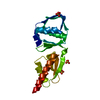
| Output model | PDB-6rwk: |