 Movie
Movie Controller


+ Open data

- Basic information
| Entry | Database: EMDB / ID: EMD-0991 | ||||||||||||
|---|---|---|---|---|---|---|---|---|---|---|---|---|---|
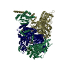
| Title | Structure of Dimethylformamidase, tetramer, E521A mutant | ||||||||||||
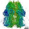
 Map data Map data | One of the half map from the refinement in Relion | ||||||||||||
 Sample Sample |
| ||||||||||||
 Keywords Keywords | ab polypeptide / mononuclear iron / amidohydrolase / tetramer / HYDROLASE | ||||||||||||
| Function / homology | N,N-dimethylformamidase / N,N-dimethylformamidase activity / N,N-dimethylformamidase beta subunit-like, C-terminal / N,N-dimethylformamidase beta subunit-like, C-terminal / Concanavalin A-like lectin/glucanase domain superfamily / metal ion binding / N,N-dimethylformamidase large subunit / N,N-dimethylformamidase small subunit Function and homology information Function and homology information | ||||||||||||
| Biological species |  Paracoccus sp. SSG05 (bacteria) Paracoccus sp. SSG05 (bacteria) | ||||||||||||
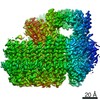
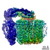
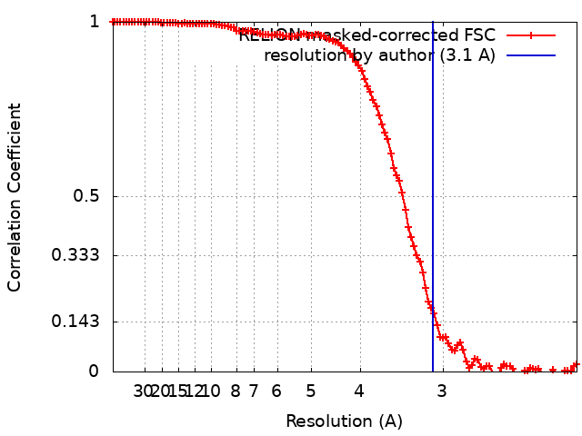
| Method | single particle reconstruction / cryo EM / Resolution: 3.1 Å | ||||||||||||
 Authors Authors | Arya CA / Yadav S | ||||||||||||
| Funding support |  India, 3 items India, 3 items
| ||||||||||||
 Citation Citation | Journal: Angew Chem Int Ed Engl / Year: 2020 Title: A 2-Tyr-1-carboxylate Mononuclear Iron Center Forms the Active Site of a Paracoccus Dimethylformamidase. Authors: Chetan Kumar Arya / Swati Yadav / Jonathan Fine / Ana Casanal / Gaurav Chopra / Gurunath Ramanathan / Kutti R Vinothkumar / Ramaswamy Subramanian /   Abstract: N,N-dimethyl formamide (DMF) is an extensively used organic solvent but is also a potent pollutant. Certain bacterial species from genera such as Paracoccus, Pseudomonas, and Alcaligenes have evolved ...N,N-dimethyl formamide (DMF) is an extensively used organic solvent but is also a potent pollutant. Certain bacterial species from genera such as Paracoccus, Pseudomonas, and Alcaligenes have evolved to use DMF as a sole carbon and nitrogen source for growth via degradation by a dimethylformamidase (DMFase). We show that DMFase from Paracoccus sp. strain DMF is a halophilic and thermostable enzyme comprising a multimeric complex of the α β or (α β ) type. One of the three domains of the large subunit and the small subunit are hitherto undescribed protein folds of unknown evolutionary origin. The active site consists of a mononuclear iron coordinated by two Tyr side-chain phenolates and one carboxylate from Glu. The Fe ion in the active site catalyzes the hydrolytic cleavage of the amide bond in DMF. Kinetic characterization reveals that the enzyme shows cooperativity between subunits, and mutagenesis and structural data provide clues to the catalytic mechanism. | ||||||||||||
| History |
|
- Structure visualization
| Movie |
Movie viewer |
|---|---|
| Structure viewer | EM map: SurfViewMolmilJmol/JSmol |
| Supplemental images |

- Downloads & links
-EMDB archive
| Map data | emd_0991.map.gz | 117 MB | EMDB map data format | |
|---|---|---|---|---|
| Header (meta data) | emd-0991-v30.xmlemd-0991.xml | 21.8 KB 21.8 KB | Display Display | EMDB header |
| FSC (resolution estimation) | emd_0991_fsc.xml | 11.4 KB | Display | FSC data file |
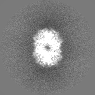
| Images | 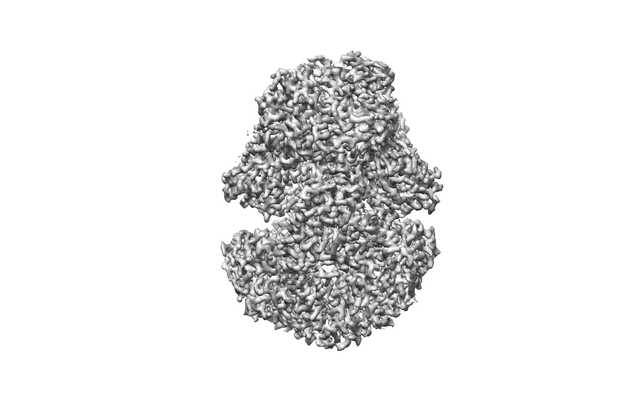 emd_0991.png emd_0991.png | 49.2 KB | ||
| Filedesc metadata | emd-0991.cif.gz | 6.9 KB | ||
| Others | emd_0991_half_map_1.map.gzemd_0991_half_map_2.map.gz | 96.7 MB 96.8 MB | ||
| Archive directory |  http://ftp.pdbj.org/pub/emdb/structures/EMD-0991ftp://ftp.pdbj.org/pub/emdb/structures/EMD-0991 http://ftp.pdbj.org/pub/emdb/structures/EMD-0991ftp://ftp.pdbj.org/pub/emdb/structures/EMD-0991 | HTTPS FTP |
-Validation report
| Summary document | emd_0991_validation.pdf.gz | 989.8 KB | Display | EMDB validaton report |
|---|---|---|---|---|
| Full document | emd_0991_full_validation.pdf.gz | 989.4 KB | Display | |
| Data in XML | emd_0991_validation.xml.gz | 18.6 KB | Display | |
| Data in CIF | emd_0991_validation.cif.gz | 24.5 KB | Display | |
| Arichive directory | https://ftp.pdbj.org/pub/emdb/validation_reports/EMD-0991ftp://ftp.pdbj.org/pub/emdb/validation_reports/EMD-0991 | HTTPS FTP |
-Related structure data
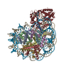
| Related structure data | 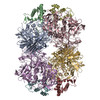 6lveMC 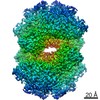 0988C  0989C  0990C  6lvbC  6lvcC 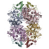 6lvdC  6lvvC C: citing same article ( M: atomic model generated by this map |
|---|---|
| Similar structure data |

-Links
| EMDB pages | EMDB (EBI/PDBe) / EMDataResource |
|---|
-Map
| File | Download / File: emd_0991.map.gz / Format: CCP4 / Size: 125 MB / Type: IMAGE STORED AS FLOATING POINT NUMBER (4 BYTES) | ||||||||||||||||||||||||||||||||||||||||||||||||||||||||||||||||||||
|---|---|---|---|---|---|---|---|---|---|---|---|---|---|---|---|---|---|---|---|---|---|---|---|---|---|---|---|---|---|---|---|---|---|---|---|---|---|---|---|---|---|---|---|---|---|---|---|---|---|---|---|---|---|---|---|---|---|---|---|---|---|---|---|---|---|---|---|---|---|
| Annotation | One of the half map from the refinement in Relion | ||||||||||||||||||||||||||||||||||||||||||||||||||||||||||||||||||||
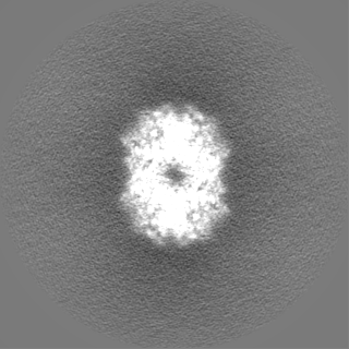
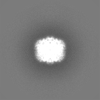
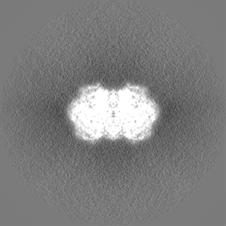
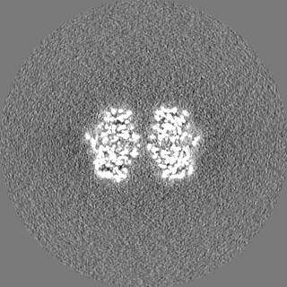
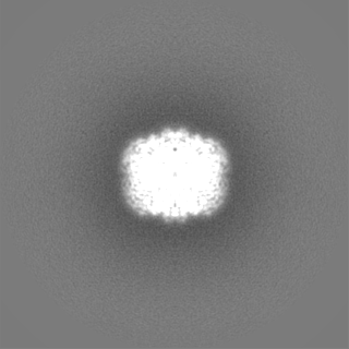
| Projections & slices | Image control
Images are generated by Spider. | ||||||||||||||||||||||||||||||||||||||||||||||||||||||||||||||||||||
| Voxel size | X=Y=Z: 1.07 Å | ||||||||||||||||||||||||||||||||||||||||||||||||||||||||||||||||||||
| Density |
| ||||||||||||||||||||||||||||||||||||||||||||||||||||||||||||||||||||
| Symmetry | Space group: 1 | ||||||||||||||||||||||||||||||||||||||||||||||||||||||||||||||||||||
| Details | EMDB XML:
CCP4 map header:
| ||||||||||||||||||||||||||||||||||||||||||||||||||||||||||||||||||||
 Z (Sec.)
 Y (Row.)
 X (Col.)


-Supplemental data
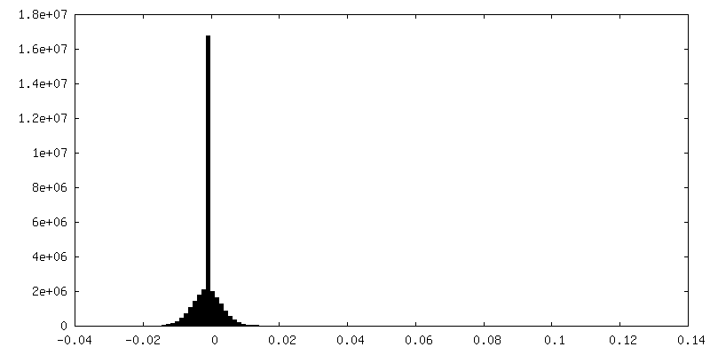
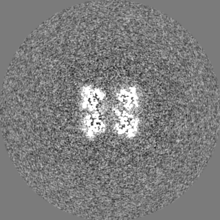
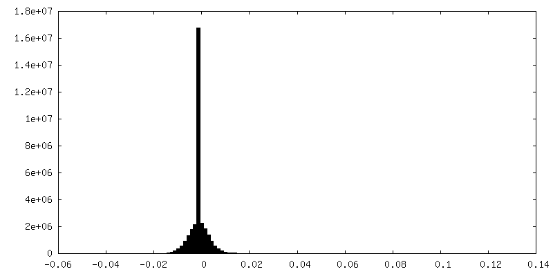
-Half map: One of the half map from the refinement in Relion
| File | emd_0991_half_map_1.map | ||||||||||||
|---|---|---|---|---|---|---|---|---|---|---|---|---|---|
| Annotation | One of the half map from the refinement in Relion | ||||||||||||
| Projections & Slices |
| ||||||||||||
| Density Histograms |


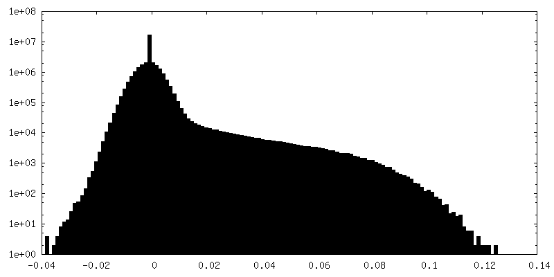
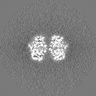
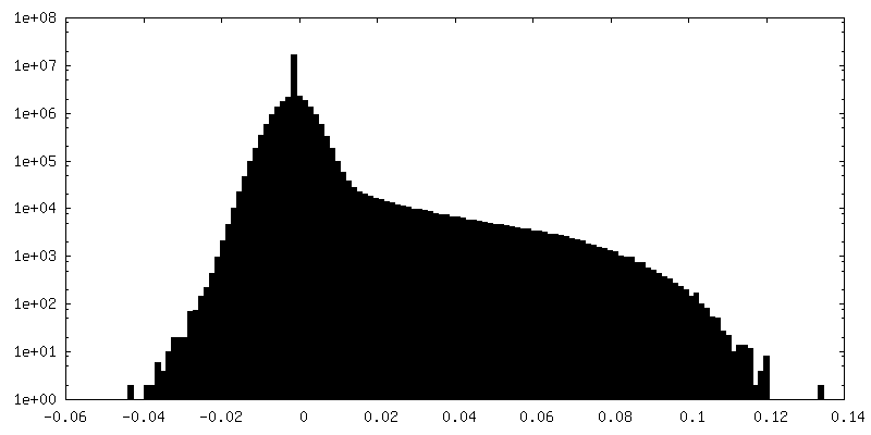
-Half map: One of the half map from the refinement in Relion
| File | emd_0991_half_map_2.map | ||||||||||||
|---|---|---|---|---|---|---|---|---|---|---|---|---|---|
| Annotation | One of the half map from the refinement in Relion | ||||||||||||
| Projections & Slices |
| ||||||||||||
| Density Histograms |


- Sample components
-Entire : Dimethylformamidase
| Entire | Name: Dimethylformamidase |
|---|---|
| Components |
|
-Supramolecule #1: Dimethylformamidase
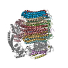
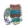
| Supramolecule | Name: Dimethylformamidase / type: complex / ID: 1 / Parent: 0 / Macromolecule list: all / Details: Tetramer, 2x(a2b2) |
|---|---|
| Source (natural) | Organism: Paracoccus sp. SSG05 (bacteria) |
| Molecular weight | Theoretical: 400 KDa |
-Macromolecule #1: N,N-dimethylformamidase large subunit
| Macromolecule | Name: N,N-dimethylformamidase large subunit / type: protein_or_peptide / ID: 1 / Number of copies: 4 / Enantiomer: LEVO / EC number: N,N-dimethylformamidase |
|---|---|
| Source (natural) | Organism: Paracoccus sp. SSG05 (bacteria) |
| Molecular weight | Theoretical: 86.283719 KDa |
| Recombinant expression | Organism: |
| Sequence | String: MKDIAIRGYC DRPSVATGET IRFYVSANET RGTFDAELVR LIHGDSNPAG PGYKEEAIKS DLEGQYPARF QRTQFGSYVE VADPDAGLQ PDGAFSVHLF LWSTTPSRGR QGIASRWNDE RQSGWNLAIE DGRVVFTIGD GSGATSSVVS DRPLFQQIWY S ITGVYDPE ...String: MKDIAIRGYC DRPSVATGET IRFYVSANET RGTFDAELVR LIHGDSNPAG PGYKEEAIKS DLEGQYPARF QRTQFGSYVE VADPDAGLQ PDGAFSVHLF LWSTTPSRGR QGIASRWNDE RQSGWNLAIE DGRVVFTIGD GSGATSSVVS DRPLFQQIWY S ITGVYDPE KKQLRLYQKS VVNRTNSRFG LVVPLDSDCA VSADATVKAA DSETSLLIAG LGEAAAQDGR TWCIAHYNGK VD APKIYGC ALGQDDAEKL SRGEIVRPIS RLAHWDFSAG IGLNGIPTDH VVDASGYGHH GRCMNQPSRG STGWNWDGHE ENF IHCPEQ YGALWFHEDC LDDCRWEKDF EFTVPEGLKS DFYAVKIRYE DTEDYIPFFV LPPRGTATAP ILVIASTLSY LAYA NEQIM HKADIGQAVA GHTPVLNEND VELHKNLSYY GLSTYDGHID GRGVQYTSWR RPIMNLRPKH RQGFGSIWEL PADLH LIDW LNHNGFEYDV ATEHDLNDQG AELLRRYKVV LTGSHPAYQT WANADAWEDY LADGGRGMYL AANGMYWIVE VHPEKP WVM EVRKELGVTA WEAPPGEYHY STNGRRGGRF RGRARATQKI WGTGMSSFGF DHSGYFVQMP DSQDERVAWI MEGIDPE ER IGDGGLVGGG AGGYELDRYD LALGTPPNTL LLASSVEHSV VYTVIPDDKA FPHPGMNGGE HPFVRADITY FSTANGGG M FATSSISWLG SLSWNDYDNN VSKMTKNVLN QFIKDEPAPR VKLAAALEHH HHHH UniProtKB: N,N-dimethylformamidase large subunit |
-Macromolecule #2: N,N-dimethylformamidase small subunit
| Macromolecule | Name: N,N-dimethylformamidase small subunit / type: protein_or_peptide / ID: 2 / Number of copies: 4 / Enantiomer: LEVO / EC number: N,N-dimethylformamidase |
|---|---|
| Source (natural) | Organism: Paracoccus sp. SSG05 (bacteria) |
| Molecular weight | Theoretical: 16.083823 KDa |
| Recombinant expression | Organism: |
| Sequence | String: MTEASESCVR DPSNYRDRSA DWYAFYDERR RKEIIDIIDE HPEIVEEHAA NPFGYRKHPS PYLQRVHNYF RMQPTFGRYY IYSEREWDA YRIATIREFG ELPELGDERF KTEEEAMHAV FLRRIEDVRA ELA UniProtKB: N,N-dimethylformamidase small subunit |
-Experimental details
-Structure determination
| Method | cryo EM |
|---|---|
 Processing Processing | single particle reconstruction |
| Aggregation state | particle |
-Sample preparation
| Concentration | 3 mg/mL | ||||||
|---|---|---|---|---|---|---|---|
| Buffer | pH: 7.2 / Component:
| ||||||
| Grid | Model: Quantifoil R0.6/1 / Material: GOLD / Mesh: 300 / Support film - Material: CARBON / Support film - topology: HOLEY / Pretreatment - Type: GLOW DISCHARGE / Pretreatment - Time: 90 sec. / Pretreatment - Atmosphere: AIR Details: Glow discharge was performed with Quorum Glocube at 25 mA for 90 seconds. | ||||||
| Vitrification | Cryogen name: ETHANE / Chamber humidity: 100 % / Chamber temperature: 291.15 K / Instrument: FEI VITROBOT MARK IV Details: Blot force was 0 and blotting was done for 3.5 seconds. | ||||||
| Details | Sample at higher salt exists mostly as tetramer |
- Electron microscopy
| Microscope | FEI TITAN KRIOS |
|---|---|
| Temperature | Min: 77.7 K / Max: 77.7 K |
| Image recording | Film or detector model: FEI FALCON III (4k x 4k) / Detector mode: COUNTING / Digitization - Dimensions - Width: 4096 pixel / Digitization - Dimensions - Height: 4096 pixel / Number grids imaged: 1 / Number real images: 1273 / Average exposure time: 60.0 sec. / Average electron dose: 27.5 e/Å2 Details: A total of 25 frames were saved from the 60 second exposure, resulting in ~1.1 electron/frame |
| Electron beam | Acceleration voltage: 300 kV / Electron source:  FIELD EMISSION GUN FIELD EMISSION GUN |
| Electron optics | C2 aperture diameter: 50.0 µm / Calibrated defocus max: 4.8126999999999995 µm / Calibrated defocus min: 1.2174 µm / Calibrated magnification: 130841 / Illumination mode: FLOOD BEAM / Imaging mode: BRIGHT FIELD / Cs: 2.7 mm / Nominal defocus max: 4.8126999999999995 µm / Nominal defocus min: 1.2174 µm / Nominal magnification: 75000 |
| Sample stage | Specimen holder model: FEI TITAN KRIOS AUTOGRID HOLDER / Cooling holder cryogen: NITROGEN |
| Experimental equipment |  Model: Titan Krios / Image courtesy: FEI Company |
+Image processing
-Atomic model buiding 1
| Refinement | Space: REAL / Protocol: OTHER / Overall B value: 50.6 |
|---|---|
| Output model | PDB-6lve: |
