 Movie
Movie Controller


+ Open data

- Basic information
| Entry | Database: EMDB / ID: EMD-0765 | |||||||||||||||
|---|---|---|---|---|---|---|---|---|---|---|---|---|---|---|---|---|
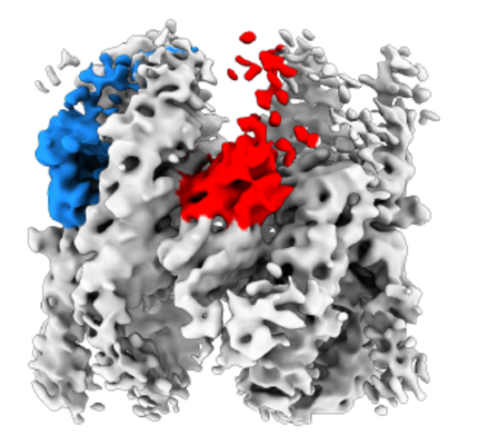
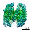
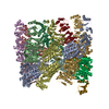
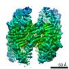
| Title | TRiC at 0.05 mM ADP-AlFx, Conformation 3, 0.05-C3 | |||||||||||||||
 Map data Map data | TRiC at 0.05 mM ADP-AlFx, Conformation 3, 0.05-C3 | |||||||||||||||
 Sample Sample |
| |||||||||||||||
| Biological species |  | |||||||||||||||
| Method | single particle reconstruction / cryo EM / Resolution: 7.18 Å | |||||||||||||||
 Authors Authors | Jin M / Cong Y | |||||||||||||||
| Funding support |  China, 4 items China, 4 items
| |||||||||||||||
 Citation Citation | Journal: Proc Natl Acad Sci U S A / Year: 2019 Title: An ensemble of cryo-EM structures of TRiC reveal its conformational landscape and subunit specificity. Authors: Mingliang Jin / Wenyu Han / Caixuan Liu / Yunxiang Zang / Jiawei Li / Fangfang Wang / Yanxing Wang / Yao Cong / Abstract: TRiC/CCT assists the folding of ∼10% of cytosolic proteins through an ATP-driven conformational cycle and is essential in maintaining protein homeostasis. Here, we determined an ensemble of cryo- ...TRiC/CCT assists the folding of ∼10% of cytosolic proteins through an ATP-driven conformational cycle and is essential in maintaining protein homeostasis. Here, we determined an ensemble of cryo-electron microscopy (cryo-EM) structures of yeast TRiC at various nucleotide concentrations, with 4 open-state maps resolved at near-atomic resolutions, and a closed-state map at atomic resolution, revealing an extra layer of an unforeseen N-terminal allosteric network. We found that, during TRiC ring closure, the CCT7 subunit moves first, responding to nucleotide binding; CCT4 is the last to bind ATP, serving as an ATP sensor; and CCT8 remains ADP-bound and is hardly involved in the ATPase-cycle in our experimental conditions; overall, yeast TRiC consumes nucleotide in a 2-ring positively coordinated manner. Our results depict a thorough picture of the TRiC conformational landscape and its allosteric transitions from the open to closed states in more structural detail and offer insights into TRiC subunit specificity in ATP consumption and ring closure, and potentially in substrate processing. | |||||||||||||||
| History |
|
- Structure visualization
| Movie |
 Movie viewer Movie viewer |
|---|---|
| Structure viewer | EM map: SurfViewMolmilJmol/JSmol |
| Supplemental images |
- Downloads & links
-EMDB archive
| Map data | emd_0765.map.gz | 26.5 MB | EMDB map data format | |
|---|---|---|---|---|
| Header (meta data) | emd-0765-v30.xmlemd-0765.xml | 10 KB 10 KB | Display Display | EMDB header |
| Images |  emd_0765.png emd_0765.png | 138 KB | ||
| Archive directory |  http://ftp.pdbj.org/pub/emdb/structures/EMD-0765ftp://ftp.pdbj.org/pub/emdb/structures/EMD-0765 http://ftp.pdbj.org/pub/emdb/structures/EMD-0765ftp://ftp.pdbj.org/pub/emdb/structures/EMD-0765 | HTTPS FTP |
-Related structure data
| Related structure data |  0756C  0757C  0758C 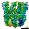 0759C 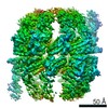 0760C 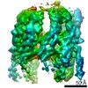 0761C 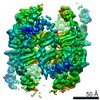 0762C 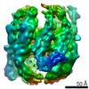 0763C 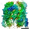 0764C 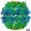 0766C 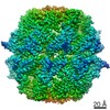 0767C  6krdC  6kreC  6ks6C 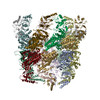 6ks7C 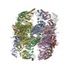 6ks8C C: citing same article ( |
|---|---|
| Similar structure data |

-Links
| EMDB pages | EMDB (EBI/PDBe) / EMDataResource |
|---|
-Map
| File | Download / File: emd_0765.map.gz / Format: CCP4 / Size: 28.7 MB / Type: IMAGE STORED AS FLOATING POINT NUMBER (4 BYTES) | ||||||||||||||||||||||||||||||||||||||||||||||||||||||||||||
|---|---|---|---|---|---|---|---|---|---|---|---|---|---|---|---|---|---|---|---|---|---|---|---|---|---|---|---|---|---|---|---|---|---|---|---|---|---|---|---|---|---|---|---|---|---|---|---|---|---|---|---|---|---|---|---|---|---|---|---|---|---|
| Annotation | TRiC at 0.05 mM ADP-AlFx, Conformation 3, 0.05-C3 | ||||||||||||||||||||||||||||||||||||||||||||||||||||||||||||
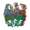
| Projections & slices | Image control
Images are generated by Spider. | ||||||||||||||||||||||||||||||||||||||||||||||||||||||||||||
| Voxel size | X=Y=Z: 1.318 Å | ||||||||||||||||||||||||||||||||||||||||||||||||||||||||||||
| Density |
| ||||||||||||||||||||||||||||||||||||||||||||||||||||||||||||
| Symmetry | Space group: 1 | ||||||||||||||||||||||||||||||||||||||||||||||||||||||||||||
| Details | EMDB XML:
CCP4 map header:
| ||||||||||||||||||||||||||||||||||||||||||||||||||||||||||||
 Z (Sec.)
 Y (Row.)
 X (Col.)


-Supplemental data
- Sample components
-Entire : TRiC complex
| Entire | Name: TRiC complex |
|---|---|
| Components |
|
-Supramolecule #1: TRiC complex
| Supramolecule | Name: TRiC complex / type: complex / ID: 1 / Parent: 0 / Macromolecule list: #1-#9 / Details: TRiC complex at 0.05 mM ADP-AlFx |
|---|---|
| Source (natural) | Organism: |
| Molecular weight | Theoretical: 960 KDa |
-Experimental details
-Structure determination
| Method | cryo EM |
|---|---|
 Processing Processing | single particle reconstruction |
| Aggregation state | particle |
-Sample preparation
| Concentration | 1 mg/mL |
|---|---|
| Buffer | pH: 7.5 |
| Vitrification | Cryogen name: ETHANE |
- Electron microscopy
| Microscope | FEI TITAN KRIOS |
|---|---|
| Image recording | Film or detector model: GATAN K2 SUMMIT (4k x 4k) / Average electron dose: 38.0 e/Å2 |
| Electron beam | Acceleration voltage: 300 kV / Electron source:  FIELD EMISSION GUN FIELD EMISSION GUN |
| Electron optics | Illumination mode: FLOOD BEAM / Imaging mode: BRIGHT FIELD |
| Experimental equipment |  Model: Titan Krios / Image courtesy: FEI Company |
-Image processing
| Final reconstruction | Resolution.type: BY AUTHOR / Resolution: 7.18 Å / Resolution method: FSC 0.143 CUT-OFF / Number images used: 24450 |
|---|---|
| Initial angle assignment | Type: MAXIMUM LIKELIHOOD |
| Final angle assignment | Type: MAXIMUM LIKELIHOOD |
