[English] 日本語
 Yorodumi
- PDB-4a0o: Symmetry-free cryo-EM map of TRiC in the nucleotide-free (apo) state -
+ Open data
- Basic information
| Entry | Database: PDB / ID: 4a0o | ||||||
|---|---|---|---|---|---|---|---|
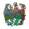
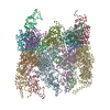
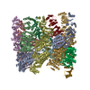
| Title | Symmetry-free cryo-EM map of TRiC in the nucleotide-free (apo) state | ||||||
 Components Components | T-COMPLEX PROTEIN 1 SUBUNIT BETA | ||||||
 Keywords Keywords | CHAPERONE / CHAPERONIN / PROTEIN FOLDING | ||||||
| Function / homology |  Function and homology information Function and homology informationAssociation of TriC/CCT with target proteins during biosynthesis / RHOBTB2 GTPase cycle / RHOBTB1 GTPase cycle / : / zona pellucida receptor complex / scaRNA localization to Cajal body / positive regulation of telomerase RNA localization to Cajal body / chaperonin-containing T-complex / binding of sperm to zona pellucida / Neutrophil degranulation ...Association of TriC/CCT with target proteins during biosynthesis / RHOBTB2 GTPase cycle / RHOBTB1 GTPase cycle / : / zona pellucida receptor complex / scaRNA localization to Cajal body / positive regulation of telomerase RNA localization to Cajal body / chaperonin-containing T-complex / binding of sperm to zona pellucida / Neutrophil degranulation / Cooperation of PDCL (PhLP1) and TRiC/CCT in G-protein beta folding / Hydrolases; Acting on acid anhydrides; In phosphorus-containing anhydrides / chaperone-mediated protein complex assembly / positive regulation of telomere maintenance via telomerase / ATP-dependent protein folding chaperone / unfolded protein binding / protein folding / cell body / microtubule / protein stabilization / ubiquitin protein ligase binding / ATP hydrolysis activity / ATP binding Similarity search - Function | ||||||
| Biological species |  | ||||||
| Method | ELECTRON MICROSCOPY / single particle reconstruction / cryo EM / Resolution: 10.5 Å | ||||||
 Authors Authors | Cong, Y. / Schroder, G.F. / Meyer, A.S. / Jakana, J. / Ma, B. / Dougherty, M.T. / Schmid, M.F. / Reissmann, S. / Levitt, M. / Ludtke, S.L. ...Cong, Y. / Schroder, G.F. / Meyer, A.S. / Jakana, J. / Ma, B. / Dougherty, M.T. / Schmid, M.F. / Reissmann, S. / Levitt, M. / Ludtke, S.L. / Frydman, J. / Chiu, W. | ||||||
 Citation Citation |  Journal: EMBO J / Year: 2012 Journal: EMBO J / Year: 2012Title: Symmetry-free cryo-EM structures of the chaperonin TRiC along its ATPase-driven conformational cycle. Authors: Yao Cong / Gunnar F Schröder / Anne S Meyer / Joanita Jakana / Boxue Ma / Matthew T Dougherty / Michael F Schmid / Stefanie Reissmann / Michael Levitt / Steven L Ludtke / Judith Frydman / Wah Chiu /  Abstract: The eukaryotic group II chaperonin TRiC/CCT is a 16-subunit complex with eight distinct but similar subunits arranged in two stacked rings. Substrate folding inside the central chamber is triggered ...The eukaryotic group II chaperonin TRiC/CCT is a 16-subunit complex with eight distinct but similar subunits arranged in two stacked rings. Substrate folding inside the central chamber is triggered by ATP hydrolysis. We present five cryo-EM structures of TRiC in apo and nucleotide-induced states without imposing symmetry during the 3D reconstruction. These structures reveal the intra- and inter-ring subunit interaction pattern changes during the ATPase cycle. In the apo state, the subunit arrangement in each ring is highly asymmetric, whereas all nucleotide-containing states tend to be more symmetrical. We identify and structurally characterize an one-ring closed intermediate induced by ATP hydrolysis wherein the closed TRiC ring exhibits an observable chamber expansion. This likely represents the physiological substrate folding state. Our structural results suggest mechanisms for inter-ring-negative cooperativity, intra-ring-positive cooperativity, and protein-folding chamber closure of TRiC. Intriguingly, these mechanisms are different from other group I and II chaperonins despite their similar architecture. | ||||||
| History |
|
- Structure visualization
| Movie |
 Movie viewer Movie viewer |
|---|---|
| Structure viewer | Molecule:  Molmil Molmil Jmol/JSmol Jmol/JSmol |
- Downloads & links
- Download
| PDBx/mmCIF format |  4a0o.cif.gz 4a0o.cif.gz | 1.2 MB | Display |  PDBx/mmCIF format PDBx/mmCIF format |
|---|---|---|---|---|
| PDB format |  pdb4a0o.ent.gz pdb4a0o.ent.gz | 997.4 KB | Display |  PDB format PDB format |
| PDBx/mmJSON format |  4a0o.json.gz 4a0o.json.gz | Tree view |  PDBx/mmJSON format PDBx/mmJSON format | |
| Others |  Other downloads Other downloads |
-Validation report
| Arichive directory |  https://data.pdbj.org/pub/pdb/validation_reports/a0/4a0o https://data.pdbj.org/pub/pdb/validation_reports/a0/4a0o ftp://data.pdbj.org/pub/pdb/validation_reports/a0/4a0o ftp://data.pdbj.org/pub/pdb/validation_reports/a0/4a0o | HTTPS FTP |
|---|
-Related structure data
| Related structure data |  1960MC  1961C  1962C  1963C  4a0vC  4a0wC  4a13C C: citing same article ( M: map data used to model this data |
|---|---|
| Similar structure data |
- Links
Links
- Assembly
| Deposited unit | 
|
|---|---|
| 1 |
|
- Components
| #1: Protein | Mass: 55107.234 Da / Num. of mol.: 16 / Fragment: RESIDUES 14-526 / Source method: isolated from a natural source / Source: (natural)  |
|---|
-Experimental details
-Experiment
| Experiment | Method: ELECTRON MICROSCOPY |
|---|---|
| EM experiment | Aggregation state: PARTICLE / 3D reconstruction method: single particle reconstruction |
- Sample preparation
| Component | Name: BOVINE TRIC IN THE NUCLEOTIDE FREE STATE / Type: COMPLEX |
|---|---|
| Specimen | Embedding applied: NO / Shadowing applied: NO / Staining applied: NO / Vitrification applied: YES |
| Specimen support | Details: HOLEY CARBON |
| Vitrification | Instrument: FEI VITROBOT MARK I / Cryogen name: ETHANE / Details: LIQUID ETHEN |
- Electron microscopy imaging
| Microscopy | Model: JEOL 3200FSC / Date: Oct 20, 2008 / Details: NONE |
|---|---|
| Electron gun | Electron source:  FIELD EMISSION GUN / Accelerating voltage: 300 kV / Illumination mode: FLOOD BEAM FIELD EMISSION GUN / Accelerating voltage: 300 kV / Illumination mode: FLOOD BEAM |
| Electron lens | Mode: BRIGHT FIELD / Nominal magnification: 50000 X / Nominal defocus max: 3000 nm / Nominal defocus min: 1200 nm |
| Specimen holder | Temperature: 101 K / Tilt angle max: 0 ° / Tilt angle min: 0 ° |
| Image recording | Electron dose: 18 e/Å2 / Film or detector model: KODAK SO-163 FILM |
| Image scans | Num. digital images: 700 |
- Processing
| EM software | Name: DireX / Category: model fitting | ||||||||||||
|---|---|---|---|---|---|---|---|---|---|---|---|---|---|
| Symmetry | Point symmetry: C1 (asymmetric) | ||||||||||||
| 3D reconstruction | Method: PROJECTION MATCHING / Resolution: 10.5 Å / Num. of particles: 47151 / Nominal pixel size: 2.4 Å / Actual pixel size: 2.4 Å Details: OUR MODELS DO NOT INCLUDE SOME REGIONS OF THE APICAL DOMAIN IN SEVERAL SUBUNITS, BECAUSE THE MAP IN THOSE REGIONS WAS NOT VERY WELL RESOLVED DUE TO THE DYNAMIC NATURE OF THOSE SUBUNITS. WE ...Details: OUR MODELS DO NOT INCLUDE SOME REGIONS OF THE APICAL DOMAIN IN SEVERAL SUBUNITS, BECAUSE THE MAP IN THOSE REGIONS WAS NOT VERY WELL RESOLVED DUE TO THE DYNAMIC NATURE OF THOSE SUBUNITS. WE FITTED THE MODEL WITH THE OCCUPANCY REFINEMENT FEATURE IN DIREX. THIS BASICALLY DETERMINES WHETHER THERE IS SUFFICIENT DENSITY FOR EACH RESIDUE OR IF THERE IS REDUCED OR MISSING DENSITY. THE OCCUPANCY IS VALUE BETWEEN 0 AND 1. WE WROTE THIS VALUE INTO THE B-FACTOR COLUMN AS 100X(1-OCCUPANCY). THIS RESEMBLES A B-FACTOR BUT IS NOT EXACTLY THE SAME. SUBMISSION BASED ON EXPERIMENTAL DATA FROM EMDB EMD-1960. (DEPOSITION ID: 10235). Symmetry type: POINT | ||||||||||||
| Refinement | Highest resolution: 10.5 Å | ||||||||||||
| Refinement step | Cycle: LAST / Highest resolution: 10.5 Å
|
 Movie
Movie Controller


 PDBj
PDBj


