Ministry of Education, Culture, Sports, Science and Technology (Japan)
Japan
Citation
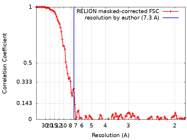
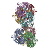
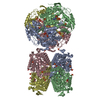
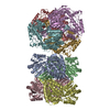
Journal: Sci Rep / Year: 2020 Title: Supramolecular tholos-like architecture constituted by archaeal proteins without functional annotation. Authors: Maho Yagi-Utsumi / Arunima Sikdar / Chihong Song / Jimin Park / Rintaro Inoue / Hiroki Watanabe / Raymond N Burton-Smith / Toshiya Kozai / Tatsuya Suzuki / Atsuji Kodama / Kentaro Ishii / ...Authors: Maho Yagi-Utsumi / Arunima Sikdar / Chihong Song / Jimin Park / Rintaro Inoue / Hiroki Watanabe / Raymond N Burton-Smith / Toshiya Kozai / Tatsuya Suzuki / Atsuji Kodama / Kentaro Ishii / Hirokazu Yagi / Tadashi Satoh / Susumu Uchiyama / Takayuki Uchihashi / Keehyoung Joo / Jooyoung Lee / Masaaki Sugiyama / Kazuyoshi Murata / Koichi Kato / Abstract: Euryarchaeal genomes encode proteasome-assembling chaperone homologs, PbaA and PbaB, although archaeal proteasome formation is a chaperone-independent process. Homotetrameric PbaB functions as a ...Euryarchaeal genomes encode proteasome-assembling chaperone homologs, PbaA and PbaB, although archaeal proteasome formation is a chaperone-independent process. Homotetrameric PbaB functions as a proteasome activator, while PbaA forms a homopentamer that does not interact with the proteasome. Notably, PbaA forms a complex with PF0014, an archaeal protein without functional annotation. In this study, based on our previous research on PbaA crystal structure, we performed an integrative analysis of the supramolecular structure of the PbaA/PF0014 complex using native mass spectrometry, solution scattering, high-speed atomic force microscopy, and electron microscopy. The results indicated that this highly thermostable complex constitutes ten PbaA and ten PF0014 molecules, which are assembled into a dumbbell-shaped structure. Two PbaA homopentameric rings correspond to the dumbbell plates, with their N-termini located outside of the plates and C-terminal segments left mobile. Furthermore, mutant PbaA lacking the mobile C-terminal segment retained the ability to form a complex with PF0014, allowing 3D modeling of the complex. The complex shows a five-column tholos-like architecture, in which each column comprises homodimeric PF0014, harboring a central cavity, which can potentially accommodate biomacromolecules including proteins. Our findings provide insight into the functional roles of Pba family proteins, offering a novel framework for designing functional protein cages.
History
Deposition
Aug 21, 2019
-
Header (metadata) release
Feb 19, 2020
-
Map release
Feb 19, 2020
-
Update
Feb 19, 2020
-
Current status
Feb 19, 2020
Processing site: PDBj / Status: Released
-
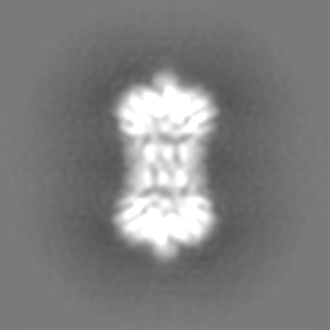
Structure visualization
Movie
Surface view with section colored by density value
In the structure databanks used in Yorodumi, some data are registered as the other names, "COVID-19 virus" and "2019-nCoV". Here are the details of the virus and the list of structure data.
Jan 31, 2019. EMDB accession codes are about to change! (news from PDBe EMDB page)
EMDB accession codes are about to change! (news from PDBe EMDB page)
The allocation of 4 digits for EMDB accession codes will soon come to an end. Whilst these codes will remain in use, new EMDB accession codes will include an additional digit and will expand incrementally as the available range of codes is exhausted. The current 4-digit format prefixed with “EMD-” (i.e. EMD-XXXX) will advance to a 5-digit format (i.e. EMD-XXXXX), and so on. It is currently estimated that the 4-digit codes will be depleted around Spring 2019, at which point the 5-digit format will come into force.
The EM Navigator/Yorodumi systems omit the EMD- prefix.
Related info.:Q: What is EMD? / ID/Accession-code notation in Yorodumi/EM Navigator
Yorodumi is a browser for structure data from EMDB, PDB, SASBDB, etc.
This page is also the successor to EM Navigator detail page, and also detail information page/front-end page for Omokage search.
The word "yorodu" (or yorozu) is an old Japanese word meaning "ten thousand". "mi" (miru) is to see.
Related info.:EMDB / PDB / SASBDB / Comparison of 3 databanks / Yorodumi Search / Aug 31, 2016. New EM Navigator & Yorodumi / Yorodumi Papers / Jmol/JSmol / Function and homology information / Changes in new EM Navigator and Yorodumi
 Movie
Movie Controller

 Yorodumi
 Open data
 Basic information
 Map data
 Sample
 Pyrococcus furiosus (archaea)
 Authors
 Japan, 1 items
  Citation

 Structure visualization
 Movie viewer
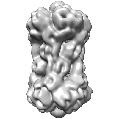
 Downloads & links
 emd_0755.png
 http://ftp.pdbj.org/pub/emdb/structures/EMD-0755

 Z (Sec.)
 Y (Row.)
 X (Col.)


 Sample components
 Processing
 Electron microscopy
 FIELD EMISSION GUN