 Movie
Movie Controller

[English] 日本語
 Yorodumi
- EMDB-0413: Vesicle-plasma interface under the docked vesicles in NGF-differe... -
+ Open data

- Basic information
| Entry | Database: EMDB / ID: EMD-0413 | |||||||||
|---|---|---|---|---|---|---|---|---|---|---|
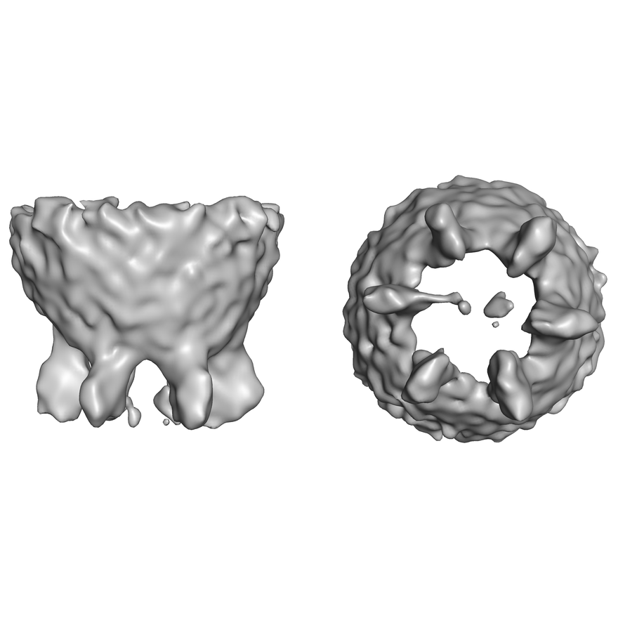
| Title | Vesicle-plasma interface under the docked vesicles in NGF-differentiated PC12 cells without imposed symmetry | |||||||||
 Map data Map data | Vesicle-plasma interface under the docked vesicles in NGF-differentiated PC12 cells without imposed symmetry | |||||||||
 Sample Sample |
| |||||||||
| Biological species |  | |||||||||
| Method | subtomogram averaging / cryo EM / Resolution: 50.0 Å | |||||||||
 Authors Authors | Li X / Radhakrishnan A / Grushin K / Krishnakumar S / Liu J / Rothman J | |||||||||
 Citation Citation | Journal: FEBS Lett / Year: 2019 Title: Symmetrical organization of proteins under docked synaptic vesicles. Authors: Xia Li / Abhijith Radhakrishnan / Kirill Grushin / Ravikiran Kasula / Arunima Chaudhuri / Sujatha Gomathinayagam / Shyam S Krishnakumar / Jun Liu / James E Rothman /    Abstract: During calcium-regulated exocytosis, the constitutive fusion machinery is 'clamped' in a partially assembled state until synchronously released by calcium. The protein machinery involved in this ...During calcium-regulated exocytosis, the constitutive fusion machinery is 'clamped' in a partially assembled state until synchronously released by calcium. The protein machinery involved in this process is known, but the supra-molecular architecture and underlying mechanisms are unclear. Here, we use cryo-electron tomography analysis in nerve growth factor-differentiated neuro-endocrine (PC12) cells to delineate the organization of the release machinery under the docked vesicles. We find that exactly six exocytosis modules, each likely consisting of a single SNAREpin with its bound Synaptotagmins, Complexin, and Munc18 proteins, are symmetrically arranged at the vesicle-PM interface. Mutational analysis suggests that the symmetrical organization is templated by circular oligomers of Synaptotagmin. The observed arrangement, including its precise radial positioning, is in-line with the recently proposed 'buttressed ring hypothesis'. | |||||||||
| History |
|
- Structure visualization
| Movie |
 Movie viewer Movie viewer |
|---|---|
| Structure viewer | EM map: SurfViewMolmilJmol/JSmol |
| Supplemental images |
- Downloads & links
-EMDB archive
| Map data | emd_0413.map.gz | 6.1 MB | EMDB map data format | |
|---|---|---|---|---|
| Header (meta data) | emd-0413-v30.xmlemd-0413.xml | 8.5 KB 8.5 KB | Display Display | EMDB header |
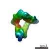
| Images |  emd_0413.png emd_0413.png | 186.2 KB | ||
| Archive directory |  http://ftp.pdbj.org/pub/emdb/structures/EMD-0413ftp://ftp.pdbj.org/pub/emdb/structures/EMD-0413 http://ftp.pdbj.org/pub/emdb/structures/EMD-0413ftp://ftp.pdbj.org/pub/emdb/structures/EMD-0413 | HTTPS FTP |
-Related structure data


-Links
| EMDB pages | EMDB (EBI/PDBe) / EMDataResource |
|---|
-Map
| File | Download / File: emd_0413.map.gz / Format: CCP4 / Size: 6.6 MB / Type: IMAGE STORED AS FLOATING POINT NUMBER (4 BYTES) | ||||||||||||||||||||||||||||||||||||||||||||||||||||||||||||
|---|---|---|---|---|---|---|---|---|---|---|---|---|---|---|---|---|---|---|---|---|---|---|---|---|---|---|---|---|---|---|---|---|---|---|---|---|---|---|---|---|---|---|---|---|---|---|---|---|---|---|---|---|---|---|---|---|---|---|---|---|---|
| Annotation | Vesicle-plasma interface under the docked vesicles in NGF-differentiated PC12 cells without imposed symmetry | ||||||||||||||||||||||||||||||||||||||||||||||||||||||||||||
| Projections & slices | Image control
Images are generated by Spider. | ||||||||||||||||||||||||||||||||||||||||||||||||||||||||||||
| Voxel size | X=Y=Z: 5.4 Å | ||||||||||||||||||||||||||||||||||||||||||||||||||||||||||||
| Density |
| ||||||||||||||||||||||||||||||||||||||||||||||||||||||||||||
| Symmetry | Space group: 1 | ||||||||||||||||||||||||||||||||||||||||||||||||||||||||||||
| Details | EMDB XML:
CCP4 map header:
| ||||||||||||||||||||||||||||||||||||||||||||||||||||||||||||
 Z (Sec.)
 Y (Row.)
 X (Col.)


-Supplemental data
- Sample components
-Entire : Docked synaptic vesicle
| Entire | Name: Docked synaptic vesicle |
|---|---|
| Components |
|
-Supramolecule #1: Docked synaptic vesicle
| Supramolecule | Name: Docked synaptic vesicle / type: organelle_or_cellular_component / ID: 1 / Parent: 0 Details: Synaptic vesicles in neurites developed from nerve growth factor (NGF)-differentiated pheochromocytoma (PC12) cells |
|---|---|
| Source (natural) | Organism: |
-Experimental details
-Structure determination
| Method | cryo EM |
|---|---|
 Processing Processing | subtomogram averaging |
| Aggregation state | cell |
-Sample preparation
| Buffer | pH: 7.4 |
|---|---|
| Grid | Model: Quantifoil R2/1 / Material: GOLD |
| Vitrification | Cryogen name: ETHANE / Instrument: HOMEMADE PLUNGER |
- Electron microscopy
| Microscope | FEI TITAN KRIOS |
|---|---|
| Image recording | Film or detector model: GATAN K2 SUMMIT (4k x 4k) / Detector mode: COUNTING / Average electron dose: 1.47 e/Å2 |
| Electron beam | Acceleration voltage: 300 kV / Electron source:  FIELD EMISSION GUN FIELD EMISSION GUN |
| Electron optics | Illumination mode: FLOOD BEAM / Imaging mode: BRIGHT FIELD |
| Experimental equipment |  Model: Titan Krios / Image courtesy: FEI Company |
-Image processing
| Final reconstruction | Applied symmetry - Point group: C1 (asymmetric) / Resolution.type: BY AUTHOR / Resolution: 50.0 Å / Resolution method: FSC 0.5 CUT-OFF / Number subtomograms used: 2434 |
|---|---|
| Extraction | Number tomograms: 574 / Number images used: 4758 |
| Final angle assignment | Type: OTHER |
