ジャーナル: FEBS Lett / 年: 2019 タイトル: Symmetrical organization of proteins under docked synaptic vesicles. 著者: Xia Li / Abhijith Radhakrishnan / Kirill Grushin / Ravikiran Kasula / Arunima Chaudhuri / Sujatha Gomathinayagam / Shyam S Krishnakumar / Jun Liu / James E Rothman / 要旨: During calcium-regulated exocytosis, the constitutive fusion machinery is 'clamped' in a partially assembled state until synchronously released by calcium. The protein machinery involved in this ...During calcium-regulated exocytosis, the constitutive fusion machinery is 'clamped' in a partially assembled state until synchronously released by calcium. The protein machinery involved in this process is known, but the supra-molecular architecture and underlying mechanisms are unclear. Here, we use cryo-electron tomography analysis in nerve growth factor-differentiated neuro-endocrine (PC12) cells to delineate the organization of the release machinery under the docked vesicles. We find that exactly six exocytosis modules, each likely consisting of a single SNAREpin with its bound Synaptotagmins, Complexin, and Munc18 proteins, are symmetrically arranged at the vesicle-PM interface. Mutational analysis suggests that the symmetrical organization is templated by circular oligomers of Synaptotagmin. The observed arrangement, including its precise radial positioning, is in-line with the recently proposed 'buttressed ring hypothesis'.
 ムービー
ムービー コントローラー


 データを開く
 基本情報
基本情報 マップデータ
 試料
試料
 Rattus norvegicus (ドブネズミ)
 データ登録者
 引用
引用


 構造の表示
構造の表示 ムービービューア
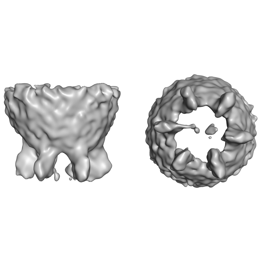
 ダウンロードとリンク
 emd_0413.png
 http://ftp.pdbj.org/pub/emdb/structures/EMD-0413

 試料の構成要素
 解析
解析 電子顕微鏡法
