[English] 日本語
 Yorodumi
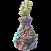
- EMDB-0150: PTC3 holotoxin complex from Photorhabdus luminiscens - Mutant TcC... -
+ Open data
- Basic information
| Entry | Database: EMDB / ID: EMD-0150 | |||||||||
|---|---|---|---|---|---|---|---|---|---|---|
| Title | PTC3 holotoxin complex from Photorhabdus luminiscens - Mutant TcC-D651A | |||||||||
 Map data Map data | CryoEM structure of the Photorhadbus luminescens ABC-D651A holotoxin complex | |||||||||
 Sample Sample |
| |||||||||
 Keywords Keywords | Tc / ABC / a-PFT / membrane transport / translocation / pore forming toxin / TOXIN | |||||||||
| Function / homology |  Function and homology information Function and homology information | |||||||||
| Biological species |  Photorhabdus luminescens (bacteria) Photorhabdus luminescens (bacteria) | |||||||||
| Method | single particle reconstruction / cryo EM / Resolution: 3.72 Å | |||||||||
 Authors Authors | Gatsogiannis C / Merino F | |||||||||
| Funding support |  Germany, 2 items Germany, 2 items
| |||||||||
 Citation Citation |  Journal: Nature / Year: 2018 Journal: Nature / Year: 2018Title: Tc toxin activation requires unfolding and refolding of a β-propeller. Authors: Christos Gatsogiannis / Felipe Merino / Daniel Roderer / David Balchin / Evelyn Schubert / Anne Kuhlee / Manajit Hayer-Hartl / Stefan Raunser /  Abstract: Tc toxins secrete toxic enzymes into host cells using a unique syringe-like injection mechanism. They are composed of three subunits, TcA, TcB and TcC. TcA forms the translocation channel and the TcB- ...Tc toxins secrete toxic enzymes into host cells using a unique syringe-like injection mechanism. They are composed of three subunits, TcA, TcB and TcC. TcA forms the translocation channel and the TcB-TcC heterodimer functions as a cocoon that shields the toxic enzyme. Binding of the cocoon to the channel triggers opening of the cocoon and translocation of the toxic enzyme into the channel. Here we show in atomic detail how the assembly of the three components activates the toxin. We find that part of the cocoon completely unfolds and refolds into an alternative conformation upon binding. The presence of the toxic enzyme inside the cocoon is essential for its subnanomolar binding affinity for the TcA subunit. The enzyme passes through a narrow negatively charged constriction site inside the cocoon, probably acting as an extruder that releases the unfolded protein with its C terminus first into the translocation channel. | |||||||||
| History |
|
- Structure visualization
| Movie |
 Movie viewer Movie viewer |
|---|---|
| Structure viewer | EM map:  SurfView SurfView Molmil Molmil Jmol/JSmol Jmol/JSmol |
| Supplemental images |
- Downloads & links
-EMDB archive
| Map data |  emd_0150.map.gz emd_0150.map.gz | 207.7 MB |  EMDB map data format EMDB map data format | |
|---|---|---|---|---|
| Header (meta data) |  emd-0150-v30.xml emd-0150-v30.xml emd-0150.xml emd-0150.xml | 19.3 KB 19.3 KB | Display Display |  EMDB header EMDB header |
| Images |  emd_0150.png emd_0150.png | 127.1 KB | ||
| Filedesc metadata |  emd-0150.cif.gz emd-0150.cif.gz | 9.1 KB | ||
| Archive directory |  http://ftp.pdbj.org/pub/emdb/structures/EMD-0150 http://ftp.pdbj.org/pub/emdb/structures/EMD-0150 ftp://ftp.pdbj.org/pub/emdb/structures/EMD-0150 ftp://ftp.pdbj.org/pub/emdb/structures/EMD-0150 | HTTPS FTP |
-Validation report
| Summary document |  emd_0150_validation.pdf.gz emd_0150_validation.pdf.gz | 215 KB | Display |  EMDB validaton report EMDB validaton report |
|---|---|---|---|---|
| Full document |  emd_0150_full_validation.pdf.gz emd_0150_full_validation.pdf.gz | 214.1 KB | Display | |
| Data in XML |  emd_0150_validation.xml.gz emd_0150_validation.xml.gz | 7.2 KB | Display | |
| Arichive directory |  https://ftp.pdbj.org/pub/emdb/validation_reports/EMD-0150 https://ftp.pdbj.org/pub/emdb/validation_reports/EMD-0150 ftp://ftp.pdbj.org/pub/emdb/validation_reports/EMD-0150 ftp://ftp.pdbj.org/pub/emdb/validation_reports/EMD-0150 | HTTPS FTP |
-Related structure data
| Related structure data |  6h6fMC  0149C  6h6eC  6h6gC M: atomic model generated by this map C: citing same article ( |
|---|---|
| Similar structure data |
- Links
Links
| EMDB pages |  EMDB (EBI/PDBe) / EMDB (EBI/PDBe) /  EMDataResource EMDataResource |
|---|---|
| Related items in Molecule of the Month |
- Map
Map
| File |  Download / File: emd_0150.map.gz / Format: CCP4 / Size: 421.9 MB / Type: IMAGE STORED AS FLOATING POINT NUMBER (4 BYTES) Download / File: emd_0150.map.gz / Format: CCP4 / Size: 421.9 MB / Type: IMAGE STORED AS FLOATING POINT NUMBER (4 BYTES) | ||||||||||||||||||||||||||||||||||||||||||||||||||||||||||||
|---|---|---|---|---|---|---|---|---|---|---|---|---|---|---|---|---|---|---|---|---|---|---|---|---|---|---|---|---|---|---|---|---|---|---|---|---|---|---|---|---|---|---|---|---|---|---|---|---|---|---|---|---|---|---|---|---|---|---|---|---|---|
| Annotation | CryoEM structure of the Photorhadbus luminescens ABC-D651A holotoxin complex | ||||||||||||||||||||||||||||||||||||||||||||||||||||||||||||
| Projections & slices | Image control
Images are generated by Spider. | ||||||||||||||||||||||||||||||||||||||||||||||||||||||||||||
| Voxel size | X=Y=Z: 1.1 Å | ||||||||||||||||||||||||||||||||||||||||||||||||||||||||||||
| Density |
| ||||||||||||||||||||||||||||||||||||||||||||||||||||||||||||
| Symmetry | Space group: 1 | ||||||||||||||||||||||||||||||||||||||||||||||||||||||||||||
| Details | EMDB XML:
CCP4 map header:
| ||||||||||||||||||||||||||||||||||||||||||||||||||||||||||||
-Supplemental data
- Sample components
-Entire : PTC3 holotoxin complex in prepore state - Mutant TcC-D651A
| Entire | Name: PTC3 holotoxin complex in prepore state - Mutant TcC-D651A |
|---|---|
| Components |
|
-Supramolecule #1: PTC3 holotoxin complex in prepore state - Mutant TcC-D651A
| Supramolecule | Name: PTC3 holotoxin complex in prepore state - Mutant TcC-D651A type: complex / ID: 1 / Parent: 0 / Macromolecule list: all Details: The catalytic protease spartyl protease site is deleted |
|---|---|
| Molecular weight | Theoretical: 1.7 MDa |
-Supramolecule #2: TcdA1
| Supramolecule | Name: TcdA1 / type: complex / ID: 2 / Parent: 1 / Macromolecule list: #1 / Details: 5 TcdA1 protomers in the PTC3 holotoxin complex |
|---|---|
| Source (natural) | Organism:  Photorhabdus luminescens (bacteria) Photorhabdus luminescens (bacteria) |
-Supramolecule #3: TcdB2/TccC3-D651A
| Supramolecule | Name: TcdB2/TccC3-D651A / type: complex / ID: 3 / Parent: 1 / Macromolecule list: #2 Details: 1 TcdB2/TccC3-D651A monomer in the PTC3 holotoxin complex. The catalytic aspartyl protease site of TccC3 is deleted. |
|---|---|
| Source (natural) | Organism:  Photorhabdus luminescens (bacteria) Photorhabdus luminescens (bacteria) |
-Macromolecule #1: TcdA1
| Macromolecule | Name: TcdA1 / type: protein_or_peptide / ID: 1 / Number of copies: 5 / Enantiomer: LEVO |
|---|---|
| Source (natural) | Organism:  Photorhabdus luminescens (bacteria) Photorhabdus luminescens (bacteria) |
| Molecular weight | Theoretical: 283.229406 KDa |
| Recombinant expression | Organism:  |
| Sequence | String: MNESVKEIPD VLKSQCGFNC LTDISHSSFN EFRQQVSEHL SWSETHDLYH DAQQAQKDNR LYEARILKRA NPQLQNAVHL AILAPNAEL IGYNNQFSGR ASQYVAPGTV SSMFSPAAYL TELYREARNL HASDSVYYLD TRRPDLKSMA LSQQNMDIEL S TLSLSNEL ...String: MNESVKEIPD VLKSQCGFNC LTDISHSSFN EFRQQVSEHL SWSETHDLYH DAQQAQKDNR LYEARILKRA NPQLQNAVHL AILAPNAEL IGYNNQFSGR ASQYVAPGTV SSMFSPAAYL TELYREARNL HASDSVYYLD TRRPDLKSMA LSQQNMDIEL S TLSLSNEL LLESIKTESK LENYTKVMEM LSTFRPSGAT PYHDAYENVR EVIQLQDPGL EQLNASPAIA GLMHQASLLG IN ASISPEL FNILTEEITE GNAEELYKKN FGNIEPASLA MPEYLKRYYN LSDEELSQFI GKASNFGQQE YSNNQLITPV VNS SDGTVK VYRITREYTT NAYQMDVELF PFGGENYRLD YKFKNFYNAS YLSIKLNDKR ELVRTEGAPQ VNIEYSANIT LNTA DISQP FEIGLTRVLP SGSWAYAAAK FTVEEYNQYS FLLKLNKAIR LSRATELSPT ILEGIVRSVN LQLDINTDVL GKVFL TKYY MQRYAIHAET ALILCNAPIS QRSYDNQPSQ FDRLFNTPLL NGQYFSTGDE EIDLNSGSTG DWRKTILKRA FNIDDV SLF RLLKITDHDN KDGKIKNNLK NLSNLYIGKL LADIHQLTID ELDLLLIAVG EGKTNLSAIS DKQLATLIRK LNTITSW LH TQKWSVFQLF IMTSTSYNKT LTPEIKNLLD TVYHGLQGFD KDKADLLHVM APYIAATLQL SSENVAHSVL LWADKLQP G DGAMTAEKFW DWLNTKYTPG SSEAVETQEH IVQYCQALAQ LEMVYHSTGI NENAFRLFVT KPEMFGAATG AAPAHDALS LIMLTRFADW VNALGEKASS VLAAFEANSL TAEQLADAMN LDANLLLQAS IQAQNHQHLP PVTPENAFSC WTSINTILQW VNVAQQLNV APQGVSALVG LDYIQSMKET PTYAQWENAA GVLTAGLNSQ QANTLHAFLD ESRSAALSTY YIRQVAKAAA A IKSRDDLY QYLLIDNQVS AAIKTTRIAE AIASIQLYVN RALENVEENA NSGVISRQFF IDWDKYNKRY STWAGVSQLV YY PENYIDP TMRIGQTKMM DALLQSVSQS QLNADTVEDA FMSYLTSFEQ VANLKVISAY HDNINNDQGL TYFIGLSETD AGE YYWRSV DHSKFNDGKF AANAWSEWHK IDCPINPYKS TIRPVIYKSR LYLLWLEQKE ITKQTGNSKD GYQTETDYRY ELKL AHIRY DGTWNTPITF DVNKKISELK LEKNRAPGLY CAGYQGEDTL LVMFYNQQDT LDSYKNASMQ GLYIFADMAS KDMTP EQSN VYRDNSYQQF DTNNVRRVNN RYAEDYEIPS SVSSRKDYGW GDYYLSMVYN GDIPTINYKA ASSDLKIYIS PKLRII HNG YEGQKRNQCN LMNKYGKLGD KFIVYTSLGV NPNNSSNKLM FYPVYQYSGN TSGLNQGRLL FHRDTTYPSK VEAWIPG AK RSLTNQNAAI GDDYATDSLN KPDDLKQYIF MTDSKGTATD VSGPVEINTA ISPAKVQIIV KAGGKEQTFT ADKDVSIQ P SPSFDEMNYQ FNALEIDGSG LNFINNSASI DVTFTAFAED GRKLGYESFS IPVTLKVSTD NALTLHHNEN GAQYMQWQS YRTRLNTLFA RQLVARATTG IDTILSMETQ NIQEPQLGKG FYATFVIPPY NLSTHGDERW FKLYIKHVVD NNSHIIYSGQ LTDTNINIT LFIPLDDVPL NQDYHAKVYM TFKKSPSDGT WWGPHFVRDD KGIVTINPKS ILTHFESVNV LNNISSEPMD F SGANSLYF WELFYYTPML VAQRLLHEQN FDEANRWLKY VWSPSGYIVH GQIQNYQWNV RPLLEDTSWN SDPLDSVDPD AV AQHDPMH YKVSTFMRTL DLLIARGDHA YRQLERDTLN EAKMWYMQAL HLLGDKPYLP LSTTWSDPRL DRAADITTQN AHD SAIVAL RQNIPTPAPL SLRSANTLTD LFLPQINEVM MNYWQTLAQR VYNLRHNLSI DGQPLYLPIY ATPADPKALL SAAV ATSQG GGKLPESFMS LWRFPHMLEN ARGMVSQLTQ FGSTLQNIIE RQDAEALNAL LQNQAAELIL TNLSIQDKTI EELDA EKTV LEKSKAGAQS RFDSYGKLYD ENINAGENQA MTLRASAAGL TTAVQASRLA GAAADLVPNI FGFAGGGSRW GAIAEA TGY VMEFSANVMN TEADKISQSE TYRRRRQEWE IQRNNAEAEL KQIDAQLKSL AVRREAAVLQ KTSLKTQQEQ TQSQLAF LQ RKFSNQALYN WLRGRLAAIY FQFYDLAVAR CLMAEQAYRW ELNDDSARFI KPGAWQGTYA GLLAGETLML SLAQMEDA H LKRDKRALEV ERTVSLAEVY AGLPKDNGPF SLAQEIDKLV SQGSGSAGSG NNNLAFGAGT DTKTSLQASV SFADLKIRE DYPASLGKIR RIKQISVTLP ALLGPYQDVQ AILSYGDKAG LANGCEALAV SHGMNDSGQF QLDFNDGKFL PFEGIAIDQG TLTLSFPNA SMPEKGKQAT MLKTLNDIIL HIRYTIK UniProtKB: TcdA1 |
-Macromolecule #2: TcdB2,TccC3,TccC3
| Macromolecule | Name: TcdB2,TccC3,TccC3 / type: protein_or_peptide / ID: 2 / Number of copies: 1 / Enantiomer: LEVO |
|---|---|
| Source (natural) | Organism:  Photorhabdus luminescens (bacteria) Photorhabdus luminescens (bacteria) |
| Molecular weight | Theoretical: 273.634656 KDa |
| Recombinant expression | Organism:  |
| Sequence | String: MQNSQDFSIT ELSLPKGGGA ITGMGEALTP TGPDGMAALS LPLPISAGRG YAPAFTLNYN SGAGNSPFGL GWDCNVMTIR RRTHFGVPH YDETDTFLGP EGEVLVVADQ PRDESTLQGI NLGATFTVTG YRSRLESHFS RLEYWQPKTT GKTDFWLIYS P DGQVHLLG ...String: MQNSQDFSIT ELSLPKGGGA ITGMGEALTP TGPDGMAALS LPLPISAGRG YAPAFTLNYN SGAGNSPFGL GWDCNVMTIR RRTHFGVPH YDETDTFLGP EGEVLVVADQ PRDESTLQGI NLGATFTVTG YRSRLESHFS RLEYWQPKTT GKTDFWLIYS P DGQVHLLG KSPQARISNP SQTTQTAQWL LEASVSSRGE QIYYQYRAED DTGCEADEIT HHLQATAQRY LHIVYYGNRT AS ETLPGLD GSAPSQADWL FYLVFDYGER SNNLKTPPAF STTGSWLCRQ DRFSRYEYGF EIRTRRLCRQ VLMYHHLQAL DSK ITEHNG PTLVSRLILN YDESAIASTL VFVRRVGHEQ DGNVVTLPPL ELAYQDFSPR HHAHWQPMDV LANFNAIQRW QLVD LKGEG LPGLLYQDKG AWWYRSAQRL GEIGSDAVTW EKMQPLSVIP SLQSNASLVD INGDGQLDWV ITGPGLRGYH SQRPD GSWT RFTPLNALPV EYTHPRAQLA DLMGAGLSDL VLIGPKSVRL YANTRDGFAK GKDVVQSGDI TLPVPGADPR KLVAFS DVL GSGQAHLVEV SATKVTCWPN LGRGRFGQPI TLPGFSQPAT EFNPAQVYLA DLDGSGPTDL IYVHTNRLDI FLNKSGN GF AEPVTLRFPE GLRFDHTCQL QMADVQGLGV ASLILSVPHM SPHHWRCDLT NMKPWLLNEM NNNMGVHHTL RYRSSSQF W LDEKAAALTT GQTPVCYLPF PIHTLWQTET EDEISGNKLV TTLRYARGAW DGREREFRGF GYVEQTDSHQ LAQGNAPER TPPALTKNWY ATGLPVIDNA LSTEYWRDDQ AFAGFSPRFT TWQDNKDVPL TPEDDNSRYW FNRALKGQLL RSELYGLDDS TNKHVPYTV TEFRSQVRRL QHTDSRYPVL WSSVVESRNY HYERIASDPQ CSQNITLSSD RFGQPLKQLS VQYPRRQQPA I NLYPDTLP DKLLANSYDD QQRQLRLTYQ QSSWHHLTNN TVRVLGLPDS TRSDIFTYGA ENVPAGGLNL ELLSDKNSLI AD DKPREYL GQQKTAYTDG QNTTPLQTPT RQALIAFTET TVFNQSTLSA FNGSIPSDKL STTLEQAGYQ QTNYLFPRTG EDK VWVAHH GYTDYGTAAQ FWRPQKQSNT QLTGKITLIW DANYCVVVQT RDAAGLTTSA KYDWRFLTPV QLTDINDNQH LITL DALGR PITLRFWGTE NGKMTGYSSP EKASFSPPSD VNAAIELKKP LPVAQCQVYA PESWMPVLSQ KTFNRLAEQD WQKLY NARI ITEDGRICTL AYRRWVQSQK AIPQLISLLN NGPRLPPHSL TLTTDRYDHD PEQQIRQQVV FSDGFGRLLQ AAARHE AGM ARQRNEDGSL IINVQHTENR WAVTGRTEYD NKGQPIRTYQ PYFLNDWRYV SNDSARQEKE AYADTHVYDP IGREIKV IT AKGWFRRTLF TPWFTVNEDE NDTAAEVKKV KMMKNIDPKL YQKTPTVSVY DNRGLIIRNI DFHRTTANGD PDTRITRH Q YDIHGHLNQS IDPRLYEAKQ TNNTIKPNFL WQYDLTGNPL CTESIDAGRT VTLNDIEGRP LLTVTATGVI QTRQYETSS LPGRLLSVAE QTPEEKTSRI TERLIWAGNT EAEKDHNLAG QCVRHYDTAG VTRLESLSLT GTVLSQSSQL LIDTQEANWT GDNETVWQN MLADDIYTTL STFDATGALL TQTDAKGNIQ RLAYDVAGQL NGSWLTLKGQ TEQVIIKSLT YSAAGQKLRE E HGNDVITE YSYEPETQRL IGIKTRRPSD TKVLQDLRYE YDPVGNVISI RNDAEATRFW HNQKVMPENT YTYDSLYQLI SA TGREMAN IGQQSHQFPS PALPSDNNTY TNYTRTYTYD RGGNLTKIQH SSPATQNNYT TNITVSNRSN RAVLSTLTED PAQ VDALFD AGGHQNTLIS GQNLNWNTRG ELQQVTLVKR DKGANDDREW YRYSGDGRRM LKINEQQASN NAQTQRVTYL PNLE LRLTQ NSTATTEDLQ VITVGEAGRA QVRVLHWESG KPEDIDNNQL RYSYDNLIGS SQLELDSEGQ IISEEEYYPY GGTAL WAAR NQTEASYKTI RYSGKERDAT GLYYYGYRYY QPWIGRWLSS APAGTIDGLN LYRMVRNNPV TLLDPDGLMP TIAERI AAL KKNKVTDSAP SPANATNVAI NIRPPVAPKP SLPKASTSSQ PTTHPIGAAN IKPTTSGSSI VAPLSPVGNK STSEISL PE SAQSSSSSTT STNLQKKSFT LYRADNRSFE EMQSKFPEGF KAWTPLDTKM ARQFASIFIG QKDTSNLPKE TVKNISTW G AKPKLKDLSN YIKYTKDKST VWVSTAINTE AGGQSSGAPL HKIDMDLYEF AIDGQKLNPL PEGRTKNMVP SLLLDTPQI ETSSIIALNH GPVNDAEISF LTTIPLKNVK PHKR UniProtKB: TcdB2, TccC3, TccC3 |
-Experimental details
-Structure determination
| Method | cryo EM |
|---|---|
 Processing Processing | single particle reconstruction |
| Aggregation state | particle |
- Sample preparation
| Buffer | pH: 8 Component:
| ||||||||
|---|---|---|---|---|---|---|---|---|---|
| Grid | Model: C-flat-2/1 / Material: COPPER / Mesh: 400 / Pretreatment - Type: GLOW DISCHARGE | ||||||||
| Vitrification | Cryogen name: ETHANE / Instrument: GATAN CRYOPLUNGE 3 |
- Electron microscopy
| Microscope | FEI TITAN KRIOS |
|---|---|
| Image recording | Film or detector model: FEI FALCON II (4k x 4k) / Number real images: 4958 / Average electron dose: 2.2 e/Å2 |
| Electron beam | Acceleration voltage: 300 kV / Electron source:  FIELD EMISSION GUN FIELD EMISSION GUN |
| Electron optics | Illumination mode: OTHER / Imaging mode: BRIGHT FIELD / Cs: 0.0 mm / Nominal magnification: 59000 |
| Sample stage | Specimen holder model: FEI TITAN KRIOS AUTOGRID HOLDER / Cooling holder cryogen: NITROGEN |
| Experimental equipment |  Model: Titan Krios / Image courtesy: FEI Company |
+ Image processing
-Atomic model buiding 1
| Refinement | Space: REAL / Protocol: FLEXIBLE FIT / Overall B value: 49.43 / Target criteria: Model to Map FSC |
|---|---|
| Output model |  PDB-6h6f: |
 Movie
Movie Controller


 Z (Sec.)
 Y (Row.)
 X (Col.)


