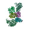
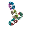
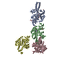
Journal: Open Biol / Year: 2018 Title: It takes two transducins to activate the cGMP-phosphodiesterase 6 in retinal rods. Authors: Bilal M Qureshi / Elmar Behrmann / Johannes Schöneberg / Justus Loerke / Jörg Bürger / Thorsten Mielke / Jan Giesebrecht / Frank Noé / Trevor D Lamb / Klaus Peter Hofmann / Christian M T ...Authors: Bilal M Qureshi / Elmar Behrmann / Johannes Schöneberg / Justus Loerke / Jörg Bürger / Thorsten Mielke / Jan Giesebrecht / Frank Noé / Trevor D Lamb / Klaus Peter Hofmann / Christian M T Spahn / Martin Heck / Abstract: Among cyclic nucleotide phosphodiesterases (PDEs), PDE6 is unique in serving as an effector enzyme in G protein-coupled signal transduction. In retinal rods and cones, PDE6 is membrane-bound and ...Among cyclic nucleotide phosphodiesterases (PDEs), PDE6 is unique in serving as an effector enzyme in G protein-coupled signal transduction. In retinal rods and cones, PDE6 is membrane-bound and activated to hydrolyse its substrate, cGMP, by binding of two active G protein α-subunits (Gα*). To investigate the activation mechanism of mammalian rod PDE6, we have collected functional and structural data, and analysed them by reaction-diffusion simulations. Gα* titration of membrane-bound PDE6 reveals a strong functional asymmetry of the enzyme with respect to the affinity of Gα* for its two binding sites on membrane-bound PDE6 and the enzymatic activity of the intermediary 1 : 1 Gα* · PDE6 complex. Employing cGMP and its 8-bromo analogue as substrates, we find that Gα* · PDE6 forms with high affinity but has virtually no cGMP hydrolytic activity. To fully activate PDE6, it takes a second copy of Gα* which binds with lower affinity, forming Gα* · PDE6 · Gα*. Reaction-diffusion simulations show that the functional asymmetry of membrane-bound PDE6 constitutes a coincidence switch and explains the lack of G protein-related noise in visual signal transduction. The high local concentration of Gα* generated by a light-activated rhodopsin molecule efficiently activates PDE6, whereas the low density of spontaneously activated Gα* fails to activate the effector enzyme.
History
Deposition
Jul 5, 2018
-
Header (metadata) release
Aug 22, 2018
-
Map release
Sep 5, 2018
-
Update
Sep 5, 2018
-
Current status
Sep 5, 2018
Processing site: PDBe / Status: Released
-
Structure visualization
Movie
Surface view with section colored by density value
In the structure databanks used in Yorodumi, some data are registered as the other names, "COVID-19 virus" and "2019-nCoV". Here are the details of the virus and the list of structure data.
Jan 31, 2019. EMDB accession codes are about to change! (news from PDBe EMDB page)
EMDB accession codes are about to change! (news from PDBe EMDB page)
The allocation of 4 digits for EMDB accession codes will soon come to an end. Whilst these codes will remain in use, new EMDB accession codes will include an additional digit and will expand incrementally as the available range of codes is exhausted. The current 4-digit format prefixed with “EMD-” (i.e. EMD-XXXX) will advance to a 5-digit format (i.e. EMD-XXXXX), and so on. It is currently estimated that the 4-digit codes will be depleted around Spring 2019, at which point the 5-digit format will come into force.
The EM Navigator/Yorodumi systems omit the EMD- prefix.
Related info.:Q: What is EMD? / ID/Accession-code notation in Yorodumi/EM Navigator
Yorodumi is a browser for structure data from EMDB, PDB, SASBDB, etc.
This page is also the successor to EM Navigator detail page, and also detail information page/front-end page for Omokage search.
The word "yorodu" (or yorozu) is an old Japanese word meaning "ten thousand". "mi" (miru) is to see.
Related info.:EMDB / PDB / SASBDB / Comparison of 3 databanks / Yorodumi Search / Aug 31, 2016. New EM Navigator & Yorodumi / Yorodumi Papers / Jmol/JSmol / Function and homology information / Changes in new EM Navigator and Yorodumi
 Movie
Movie Controller


 Open data
 Basic information
 Map data
 Sample
 Authors
 Citation

 Structure visualization
 Movie viewer
 UCSF Chimera
 Downloads & links
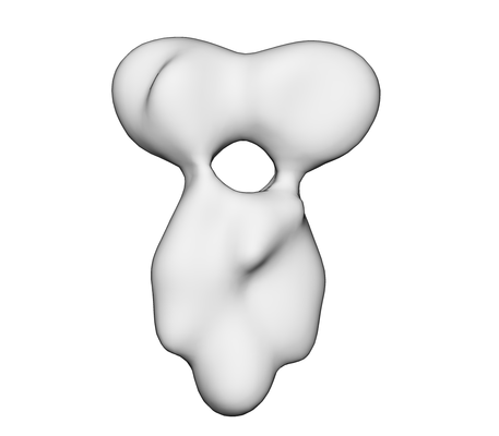 emd_0102.png
 http://ftp.pdbj.org/pub/emdb/structures/EMD-0102


 Z (Sec.)
 Y (Row.)
 X (Col.)


 Sample components
 Processing
 Electron microscopy
