 Movie
Movie Controller

[English] 日本語
 Yorodumi
- EMDB-0020: Hexameric cytochrome c nitrite reductase from the bacterium Thioa... -
+ Open data

- Basic information
| Entry |  | |||||||||
|---|---|---|---|---|---|---|---|---|---|---|
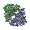
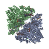
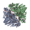
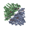
| Title | Hexameric cytochrome c nitrite reductase from the bacterium Thioalkalivibrio nitratireducens (TvNiR) | |||||||||
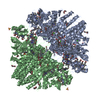
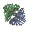
 Map data Map data | Cytochrome c nitrite reductase from the bacterium Thioalkalivibrio nitratireducens (TvNiR). | |||||||||
 Sample Sample |
| |||||||||
| Biological species |  Thioalkalivibrio nitratireducens (bacteria) Thioalkalivibrio nitratireducens (bacteria) | |||||||||
| Method | single particle reconstruction / cryo EM / Resolution: 2.56 Å | |||||||||
 Authors Authors | Baymukhametov TN / Chesnokov YM / Pichkur EB / Boyko KM / Tikhonova TV / Myasnikov AG / Vasiliev AL / Lipkin AV / Popov VO / Kovalchuk MV | |||||||||
 Citation Citation | Journal: Acta Naturae / Year: 2018 Title: Three-Dimensional Structure of Cytochrome c Nitrite Reductase As Determined by Cryo-Electron Microscopy. Authors: T N Baymukhametov / Y M Chesnokov / E B Pichkur / K M Boyko / T V Tikhonova / A G Myasnikov / A L Vasiliev / A V Lipkin / V O Popov / M V Kovalchuk /    Abstract: The structure of cytochrome c nitrite reductase from the bacterium Thioalkalivibrio nitratireducens was determined by cryo-electron microscopy (cryo-EM) at a 2.56 Å resolution. Possible structural ...The structure of cytochrome c nitrite reductase from the bacterium Thioalkalivibrio nitratireducens was determined by cryo-electron microscopy (cryo-EM) at a 2.56 Å resolution. Possible structural heterogeneity of the enzyme was assessed. The backbone and side-chain orientations in the cryo-EM-based model are, in general, similar to those in the high-resolution X-ray diffraction structure of this enzyme. | |||||||||
| History |
|
- Structure visualization
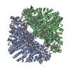
| Structure viewer | EM map:  SurfViewMolmilJmol/JSmol SurfViewMolmilJmol/JSmol |
|---|---|
| Supplemental images |

- Downloads & links
-EMDB archive
| Map data | emd_0020.map.gz | 8.7 MB | EMDB map data format | |
|---|---|---|---|---|
| Header (meta data) | emd-0020-v30.xmlemd-0020.xml | 12.1 KB 12.1 KB | Display Display | EMDB header |
| Images | 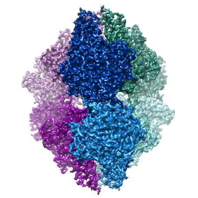 emd_0020.png emd_0020.png | 211.8 KB | ||
| Archive directory |  http://ftp.pdbj.org/pub/emdb/structures/EMD-0020ftp://ftp.pdbj.org/pub/emdb/structures/EMD-0020 http://ftp.pdbj.org/pub/emdb/structures/EMD-0020ftp://ftp.pdbj.org/pub/emdb/structures/EMD-0020 | HTTPS FTP |
-Validation report
| Summary document | emd_0020_validation.pdf.gz | 235.8 KB | Display | EMDB validaton report |
|---|---|---|---|---|
| Full document | emd_0020_full_validation.pdf.gz | 234.9 KB | Display | |
| Data in XML | emd_0020_validation.xml.gz | 6.3 KB | Display | |
| Arichive directory | https://ftp.pdbj.org/pub/emdb/validation_reports/EMD-0020ftp://ftp.pdbj.org/pub/emdb/validation_reports/EMD-0020 | HTTPS FTP |
-Related structure data
| Similar structure data |
|---|


-Links
| EMDB pages | EMDB (EBI/PDBe) / EMDataResource |
|---|
-Map
| File | Download / File: emd_0020.map.gz / Format: CCP4 / Size: 64 MB / Type: IMAGE STORED AS FLOATING POINT NUMBER (4 BYTES) | ||||||||||||||||||||||||||||||||||||
|---|---|---|---|---|---|---|---|---|---|---|---|---|---|---|---|---|---|---|---|---|---|---|---|---|---|---|---|---|---|---|---|---|---|---|---|---|---|
| Annotation | Cytochrome c nitrite reductase from the bacterium Thioalkalivibrio nitratireducens (TvNiR). | ||||||||||||||||||||||||||||||||||||
| Projections & slices | Image control
Images are generated by Spider. | ||||||||||||||||||||||||||||||||||||
| Voxel size | X=Y=Z: 0.86 Å | ||||||||||||||||||||||||||||||||||||
| Density |
| ||||||||||||||||||||||||||||||||||||
| Symmetry | Space group: 1 | ||||||||||||||||||||||||||||||||||||
| Details | EMDB XML:
|
 Z (Sec.)
 Y (Row.)
 X (Col.)


-Supplemental data
- Sample components
-Entire : Cytochrome c nitrite reductase from the bacterium Thioalkalivibri...
| Entire | Name: Cytochrome c nitrite reductase from the bacterium Thioalkalivibrio nitratireducens (TvNiR) |
|---|---|
| Components |
|
-Supramolecule #1: Cytochrome c nitrite reductase from the bacterium Thioalkalivibri...
| Supramolecule | Name: Cytochrome c nitrite reductase from the bacterium Thioalkalivibrio nitratireducens (TvNiR) type: complex / ID: 1 / Parent: 0 / Macromolecule list: all / Details: Hexameric form |
|---|---|
| Source (natural) | Organism: Thioalkalivibrio nitratireducens (bacteria) |
-Macromolecule #1: cytochrome c nitrite reductase
| Macromolecule | Name: cytochrome c nitrite reductase / type: protein_or_peptide / ID: 1 / Enantiomer: LEVO / EC number: nitrite reductase (cytochrome; ammonia-forming) |
|---|---|
| Source (natural) | Organism: Thioalkalivibrio nitratireducens (bacteria) |
| Sequence | String: MNDLNRLGRV GRWIAGAACL FLASAAHAEP GENLKPVDAM QCFDCHTQIE DMHTVGKHAT VNCVHCHDAT EHVETASSRR MGERPVTRMD LEACATCHTA QFNSFVEVRH ESHPRLEKAT PTSRSPMFDK LIAGHGFAFE HAEPRSHAFM LVDHFVVDRA YGGRFQFKNW ...String: MNDLNRLGRV GRWIAGAACL FLASAAHAEP GENLKPVDAM QCFDCHTQIE DMHTVGKHAT VNCVHCHDAT EHVETASSRR MGERPVTRMD LEACATCHTA QFNSFVEVRH ESHPRLEKAT PTSRSPMFDK LIAGHGFAFE HAEPRSHAFM LVDHFVVDRA YGGRFQFKNW QKVTDGMGAV RGAWTVLTDA DPESSDQRRF LSQTATAANP VCLNCKTQDH ILDWAYMGDE HEAAKWSRTS EVVEFARDLN HPLNCFMCHD PHSAGPRVVR DGLINAVVDR GLGTYPHDPV KSEQQGMTKV TFQRGREDFR AIGLLDTADS NVMCAQCHVE YNCNPGYQLS DGSRVGMDDR RANHFFWANV FDYKEAAQEI DFFDFRHATT GAALPKLQHP EAETFWGSVH ERNGVACADC HMPKVQLENG KVYTSHSQRT PRDMMGQACL NCHAEWTEDQ ALYAIDYIKN YTHGKIVKSE YWLAKMIDLF PVAKRAGVSE DVLNQARELH YDAHLYWEWW TAENSVGFHN PDQARESLMT SISKSKEAVS LLNDAIDAQV ASR |
-Experimental details
-Structure determination
| Method | cryo EM |
|---|---|
 Processing Processing | single particle reconstruction |
| Aggregation state | particle |
-Sample preparation
| Concentration | 6 mg/mL |
|---|---|
| Buffer | pH: 7 |
| Grid | Model: Quantifoil R1.2/1.3 / Material: COPPER / Mesh: 300 / Support film - Material: CARBON / Support film - topology: HOLEY ARRAY / Pretreatment - Type: GLOW DISCHARGE / Pretreatment - Atmosphere: AIR / Pretreatment - Pressure: 0.026 kPa |
| Vitrification | Cryogen name: ETHANE / Chamber humidity: 98 % / Chamber temperature: 277 K / Instrument: FEI VITROBOT MARK IV |
- Electron microscopy
| Microscope | FEI TITAN KRIOS |
|---|---|
| Image recording | Film or detector model: FEI FALCON II (4k x 4k) / Detector mode: INTEGRATING / Digitization - Dimensions - Width: 4096 pixel / Digitization - Dimensions - Height: 4096 pixel / Digitization - Frames/image: 1-30 / Number real images: 3055 / Average electron dose: 3.3 e/Å2 |
| Electron beam | Acceleration voltage: 300 kV / Electron source:  FIELD EMISSION GUN FIELD EMISSION GUN |
| Electron optics | Illumination mode: SPOT SCAN / Imaging mode: BRIGHT FIELD / Nominal magnification: 75000 |
| Sample stage | Specimen holder model: FEI TITAN KRIOS AUTOGRID HOLDER |
| Experimental equipment |  Model: Titan Krios / Image courtesy: FEI Company |
-Image processing
| CTF correction | Software - Name: Gctf (ver. 1.18) |
|---|---|
| Final reconstruction | Applied symmetry - Point group: D3 (2x3 fold dihedral) / Resolution.type: BY AUTHOR / Resolution: 2.56 Å / Resolution method: FSC 0.143 CUT-OFF / Software - Name: RELION (ver. 2.1) / Number images used: 33891 |
| Initial angle assignment | Type: MAXIMUM LIKELIHOOD / Software - Name: RELION (ver. 2.1) |
| Final angle assignment | Type: MAXIMUM LIKELIHOOD / Software - Name: RELION (ver. 2.1) |
-Atomic model buiding 1
| Refinement | Space: REAL / Protocol: RIGID BODY FIT |
|---|
