 Movie
Movie Controller
[English] 日本語
 Yorodumi
- SASDDP5: Mutant mammalian prion protein mRNA (octo-repear PrP mRNA) with K... -
+ Open data

- Basic information
| Entry | Database: SASBDB / ID: SASDDP5 |
|---|---|
 Sample Sample | Mutant mammalian prion protein mRNA (octo-repear PrP mRNA) with KCl and pyridostatin (PDS)
|
| Biological species | human PrP ORF |
 Citation Citation | Journal: Sci Rep / Year: 2019 Title: Octa-repeat domain of the mammalian prion protein mRNA forms stable A-helical hairpin structure rather than G-quadruplexes. Authors: Andreas Czech / Petr V Konarev / Ingrid Goebel / Dmitri I Svergun / Peter R Wills / Zoya Ignatova /    Abstract: Misfolding and aggregation of prion protein (PrP) causes neurodegenerative diseases like Creutzfeldt-Jakob disease (CJD) and scrapie. Besides the consensus that spontaneous conversion of normal ...Misfolding and aggregation of prion protein (PrP) causes neurodegenerative diseases like Creutzfeldt-Jakob disease (CJD) and scrapie. Besides the consensus that spontaneous conversion of normal cellular PrP into misfolded and aggregating PrP is the central event in prion disease, an alternative hypothesis suggests the generation of pathological PrP by rare translational frameshifting events in the octa-repeat domain of the PrP mRNA. Ribosomal frameshifting most commonly relies on a slippery site and an adjacent stable RNA structure to stall translating ribosome. Hence, it is crucial to unravel the secondary structure of the octa-repeat domain of PrP mRNA. Each of the five octa-repeats contains a motif (GGCGGUGGUGGCUGGG) which alone in vitro forms a G-quadruplex. Since the propensity of mRNA to form secondary structure depends on the sequence context, we set to determine the structure of the complete octa-repeat region. We assessed the structure of full-length octa-repeat domain of PrP mRNA using dynamic light scattering (DLS), small angle X-ray scattering (SAXS), circular dichroism (CD) spectroscopy and selective 2'-hydroxyl acylation analysis by primer extension (SHAPE). Our data show that the PrP octa-repeat mRNA forms stable A-helical hairpins with no evidence of G-quadruplex structure even in the presence of G-quadruplex stabilizing agents. |
 Contact author Contact author |
|
- Structure visualization
| Structure viewer | Molecule:  MolmilJmol/JSmol MolmilJmol/JSmol |
|---|
- Downloads & links
 SASDDP5


-Models
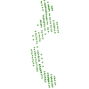
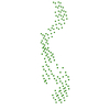
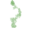
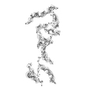
| Model #1898 |   Type: dummy / Software: (5.0) / Radius of dummy atoms: 7.75 A / Chi-square value: 1.131 / P-value: 0.000730  Search similar-shape structures of this assembly by Omokage search (details) Search similar-shape structures of this assembly by Omokage search (details) |
|---|
-Sample
| Sample | Name: Mutant mammalian prion protein mRNA (octo-repear PrP mRNA) with KCl and pyridostatin (PDS) Specimen concentration: 0.50-4.00 |
|---|---|
| Buffer | Name: 10 mM Tris buffer with 100 mM KCl and 1 mM PDS / pH: 7.5 |
| Entity #1022 | Name: PrPmRNA_mut / Type: RNA / Description: octo-repeat PrP mRNA mutant / Formula weight: 71.843 / Num. of mol.: 2 / Source: human PrP ORF Sequence: AACACUGGGG GCAGCCGAUA CCCGGGGCAG GGCAGCCCUG GAGGCAACCG CUACCCACCU CAGGGCGCUG CUGCCUGGGG GCAGCCUCAU GCUGCUGCCU GGGGGCAGCC UCAUGCUGCU GCCUGGGGGC AGCCCCAUGC UGCUGCCUGG GGACAGCCUC AUGCUGCUGC ...Sequence: AACACUGGGG GCAGCCGAUA CCCGGGGCAG GGCAGCCCUG GAGGCAACCG CUACCCACCU CAGGGCGCUG CUGCCUGGGG GCAGCCUCAU GCUGCUGCCU GGGGGCAGCC UCAUGCUGCU GCCUGGGGGC AGCCCCAUGC UGCUGCCUGG GGACAGCCUC AUGCUGCUGC CUGGGGUCAA GGAGGUGGCA CCCACCUCAG GGCGCUGCUG CCUGGGGGCA GC |
-Experimental information
| Beam | Instrument name: PETRA III EMBL P12 / City: Hamburg / 国: Germany / Type of source: X-ray synchrotron / Wavelength: 0.124 Å / Dist. spec. to detc.: 3.1 mm | ||||||||||||||||||||||||||||||||||||||||||
|---|---|---|---|---|---|---|---|---|---|---|---|---|---|---|---|---|---|---|---|---|---|---|---|---|---|---|---|---|---|---|---|---|---|---|---|---|---|---|---|---|---|---|---|
| Detector | Name: Pilatus 2M | ||||||||||||||||||||||||||||||||||||||||||
| Scan |  Measurement date: Jun 6, 2017 / Storage temperature: 20 °C / Cell temperature: 20 °C / Exposure time: 0.05 sec. / Number of frames: 20 / Unit: 1/nm /
| ||||||||||||||||||||||||||||||||||||||||||
| Distance distribution function P(R) | 
| ||||||||||||||||||||||||||||||||||||||||||
| Result | 
|
