- SASDDH5: Mammalian prion protein mRNA (PrP mRNA wild type) with KCl -
+
Open data
ID or keywords:
Loading...
-
Basic information
Entry
Database: SASBDB / ID: SASDDH5
Sample
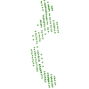
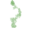
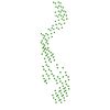
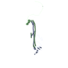
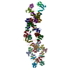
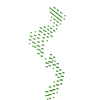
Mammalian prion protein mRNA (PrP mRNA wild type) with KCl
octo-repeat PrP mRNA (RNA), PrPmRNA, human PrP ORF
Biological species
human PrP ORF
Citation
Journal: Sci Rep / Year: 2019 Title: Octa-repeat domain of the mammalian prion protein mRNA forms stable A-helical hairpin structure rather than G-quadruplexes. Authors: Andreas Czech / Petr V Konarev / Ingrid Goebel / Dmitri I Svergun / Peter R Wills / Zoya Ignatova / Abstract: Misfolding and aggregation of prion protein (PrP) causes neurodegenerative diseases like Creutzfeldt-Jakob disease (CJD) and scrapie. Besides the consensus that spontaneous conversion of normal ...Misfolding and aggregation of prion protein (PrP) causes neurodegenerative diseases like Creutzfeldt-Jakob disease (CJD) and scrapie. Besides the consensus that spontaneous conversion of normal cellular PrP into misfolded and aggregating PrP is the central event in prion disease, an alternative hypothesis suggests the generation of pathological PrP by rare translational frameshifting events in the octa-repeat domain of the PrP mRNA. Ribosomal frameshifting most commonly relies on a slippery site and an adjacent stable RNA structure to stall translating ribosome. Hence, it is crucial to unravel the secondary structure of the octa-repeat domain of PrP mRNA. Each of the five octa-repeats contains a motif (GGCGGUGGUGGCUGGG) which alone in vitro forms a G-quadruplex. Since the propensity of mRNA to form secondary structure depends on the sequence context, we set to determine the structure of the complete octa-repeat region. We assessed the structure of full-length octa-repeat domain of PrP mRNA using dynamic light scattering (DLS), small angle X-ray scattering (SAXS), circular dichroism (CD) spectroscopy and selective 2'-hydroxyl acylation analysis by primer extension (SHAPE). Our data show that the PrP octa-repeat mRNA forms stable A-helical hairpins with no evidence of G-quadruplex structure even in the presence of G-quadruplex stabilizing agents.
Contact author
Petr Konarev (EMBL-Hamburg, European Molecular Biology Laboratory (EMBL) - Hamburg outstation, Notkestraße 85, Geb. 25A, 22607 Hamburg, Deutschland, Germany)
Instrument name: PETRA III EMBL P12 / City: Hamburg / 国: Germany / Type of source: X-ray synchrotron / Wavelength: 0.124 Å / Dist. spec. to detc.: 3.1 mm
Detector
Name: Pilatus 2M
Scan
Title: Mammalian prion protein mRNA (PrP mRNA wild type) with KCl Measurement date: Jun 6, 2017 / Storage temperature: 20 °C / Cell temperature: 20 °C / Exposure time: 0.05 sec. / Number of frames: 20 / Unit: 1/nm /
Min
Max
Q
0.1161
5.0327
Distance distribution function P(R)
Sofotware P(R): GNOM 4.6 / Number of points: 400 /
Min
Max
Q
0.1189
3.41
P(R) point
1
400
R
0
31
Result
Type of curve: merged
Experimental
Standard
Standard error
Porod
MW
180 kDa
180 kDa
20
220 kDa
Volume
-
-
-
240 nm3
P(R)
Guinier
Guinier error
P(R) error
Forward scattering, I0
123800
122435
1243
-
Radius of gyration, Rg
9.3 nm
8.81 nm
0.09
0.1
Min
Max
Error
D
-
31
1
Guinier point
1
14
-
+
About Yorodumi
-
News
-
Feb 9, 2022. New format data for meta-information of EMDB entries
New format data for meta-information of EMDB entries
Version 3 of the EMDB header file is now the official format.
The previous official version 1.9 will be removed from the archive.
In the structure databanks used in Yorodumi, some data are registered as the other names, "COVID-19 virus" and "2019-nCoV". Here are the details of the virus and the list of structure data.
Jan 31, 2019. EMDB accession codes are about to change! (news from PDBe EMDB page)
EMDB accession codes are about to change! (news from PDBe EMDB page)
The allocation of 4 digits for EMDB accession codes will soon come to an end. Whilst these codes will remain in use, new EMDB accession codes will include an additional digit and will expand incrementally as the available range of codes is exhausted. The current 4-digit format prefixed with “EMD-” (i.e. EMD-XXXX) will advance to a 5-digit format (i.e. EMD-XXXXX), and so on. It is currently estimated that the 4-digit codes will be depleted around Spring 2019, at which point the 5-digit format will come into force.
The EM Navigator/Yorodumi systems omit the EMD- prefix.
Related info.:Q: What is EMD? / ID/Accession-code notation in Yorodumi/EM Navigator
Yorodumi is a browser for structure data from EMDB, PDB, SASBDB, etc.
This page is also the successor to EM Navigator detail page, and also detail information page/front-end page for Omokage search.
The word "yorodu" (or yorozu) is an old Japanese word meaning "ten thousand". "mi" (miru) is to see.
Related info.:EMDB / PDB / SASBDB / Comparison of 3 databanks / Yorodumi Search / Aug 31, 2016. New EM Navigator & Yorodumi / Yorodumi Papers / Jmol/JSmol / Function and homology information / Changes in new EM Navigator and Yorodumi
 Movie
Movie Controller

 Open data
 Basic information
 Sample
 Citation


 Contact author
 Structure visualization
 Molmil
 Downloads & links
 SASDDH5


 Search similar-shape structures of this assembly by Omokage search (details)


