 ムービー
ムービー コントローラー

+ データを開く

- 基本情報
基本情報
| 登録情報 | データベース: SASBDB / ID: SASDD88 |
|---|---|
 試料 試料 | The BRCT domain from Mycobacterium tuberculosis DNA ligase
|
| 機能・相同性 |  機能・相同性情報 機能・相同性情報DNA ligase (NAD+) / DNA ligase (NAD+) activity / peptidoglycan-based cell wall / DNA replication / DNA repair / magnesium ion binding / plasma membrane / cytosol 類似検索 - 分子機能 |
| 生物種 |   Mycobacterium tuberculosis (結核菌) Mycobacterium tuberculosis (結核菌) |
 登録者 登録者 |
|
- 構造の表示
構造の表示
| 構造ビューア | 分子: MolmilJmol/JSmol |
|---|
- ダウンロードとリンク
 SASDD88
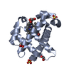


-モデル
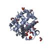
| モデル #2056 |  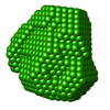 タイプ: dummy / ダミー原子の半径: 1.30 A / 対称性: P1 / カイ2乗値: 0.785 / P-value: 0.000494  Omokage検索でこの集合体の類似形状データを探す (詳細) Omokage検索でこの集合体の類似形状データを探す (詳細) |
|---|---|
| モデル #2058 |  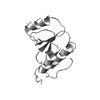 タイプ: atomic / 対称性: P1 / カイ2乗値: 4.49835837712976 Omokage検索でこの集合体の類似形状データを探す (詳細) |
| モデル #2091 |  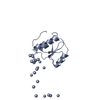 タイプ: atomic / ダミー原子の半径: 1.90 A / カイ2乗値: 2.79002473758130 Omokage検索でこの集合体の類似形状データを探す (詳細) |
-試料
| 試料 | 名称: The BRCT domain from Mycobacterium tuberculosis DNA ligase 試料濃度: 10 mg/ml |
|---|---|
| バッファ | 名称: 50 mM Tris-HCl 500 mM NaCl 5mM β-mercaptoethanol / pH: 8 |
| 要素 #1092 | 名称: BRCT / タイプ: protein / 記述: M.tb. LigA BRCT domain (DNA ligase A) / 分子量: 9.449 / 分子数: 1 / 由来: Mycobacterium tuberculosis / 参照: UniProt: P9WNV1 配列: VDERDESVPR TLAGLTIVVT GSLTGFSRDD AKEAIVARGG KAAGSVSKKT NYVVAGDSPG SKYDKAVELG VPILDEDGFR RLLADGPASR T |
-実験情報
| ビーム | 設備名称: CSIR-Central Drug Research Institute Anton Paar SAXSpace 地域: Lucknow / 国: India  / 線源: X-ray in house / 波長: 0.154 Å / スペクトロメータ・検出器間距離: 0.3171 mm / 線源: X-ray in house / 波長: 0.154 Å / スペクトロメータ・検出器間距離: 0.3171 mm | ||||||||||||||||||||||||||||||
|---|---|---|---|---|---|---|---|---|---|---|---|---|---|---|---|---|---|---|---|---|---|---|---|---|---|---|---|---|---|---|---|
| 検出器 | 名称: Mythen2 R 1K / タイプ: Hybrid Photon Counting (HPC) | ||||||||||||||||||||||||||||||
| スキャン |  測定日: 2018年6月2日 / 保管温度: 10 °C / セル温度: 10 °C / 照射時間: 1800 sec. / フレーム数: 2 / 単位: 1/nm /
| ||||||||||||||||||||||||||||||
| 距離分布関数 P(R) | 
| ||||||||||||||||||||||||||||||
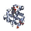
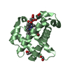
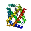
| 結果 |  コメント: The experimental molecular weight quoted for this entry was evaluated using calibrated size-exclusion chromatography (GE Healthcare Superdex75 10/30 column). The atomistic models ...コメント: The experimental molecular weight quoted for this entry was evaluated using calibrated size-exclusion chromatography (GE Healthcare Superdex75 10/30 column). The atomistic models displayed in this entry (ribbon format) are a parent Phyre2 protein homology model (top) and one of ten structures obtained from the parent after elNémo normal mode calculations (bottom). Refer to: Kelley et al. (2015) Nature Protocols 10, 845-858; Suhre & Sanejouand (2004) Nucleic Acids Res. 32(Web Server issue): W610–W614.
|
