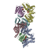
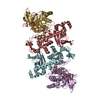
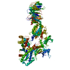
+ データを開く
- 基本情報
基本情報
| 登録情報 | データベース: SASBDB / ID: SASDCN3 |
|---|---|
 試料 試料 | Phosphoenolpyruvate-protein phosphotransferase
|
| 機能・相同性 |  機能・相同性情報 機能・相同性情報phosphoenolpyruvate-protein phosphotransferase / phosphoenolpyruvate-protein phosphotransferase activity / phosphoenolpyruvate-dependent sugar phosphotransferase system / kinase activity / metal ion binding / cytoplasm 類似検索 - 分子機能 |
| 生物種 |  |
 引用 引用 |  ジャーナル: J Am Chem Soc / 年: 2010 ジャーナル: J Am Chem Soc / 年: 2010タイトル: Solution structure of the 128 kDa enzyme I dimer from Escherichia coli and its 146 kDa complex with HPr using residual dipolar couplings and small- and wide-angle X-ray scattering. 著者: Charles D Schwieters / Jeong-Yong Suh / Alexander Grishaev / Rodolfo Ghirlando / Yuki Takayama / G Marius Clore /  要旨: The solution structures of free Enzyme I (EI, ∼128 kDa, 575 × 2 residues), the first enzyme in the bacterial phosphotransferase system, and its complex with HPr (∼146 kDa) have been solved using ...The solution structures of free Enzyme I (EI, ∼128 kDa, 575 × 2 residues), the first enzyme in the bacterial phosphotransferase system, and its complex with HPr (∼146 kDa) have been solved using novel methodology that makes use of prior structural knowledge (namely, the structures of the dimeric EIC domain and the isolated EIN domain both free and complexed to HPr), combined with residual dipolar coupling (RDC), small- (SAXS) and wide- (WAXS) angle X-ray scattering and small-angle neutron scattering (SANS) data. The calculational strategy employs conjoined rigid body/torsion/Cartesian simulated annealing, and incorporates improvements in calculating and refining against SAXS/WAXS data that take into account complex molecular shapes in the description of the solvent layer resulting in a better representation of the SAXS/WAXS data. The RDC data orient the symmetrically related EIN domains relative to the C(2) symmetry axis of the EIC dimer, while translational, shape, and size information is provided by SAXS/WAXS. The resulting structures are independently validated by SANS. Comparison of the structures of the free EI and the EI-HPr complex with that of the crystal structure of a trapped phosphorylated EI intermediate reveals large (∼70-90°) hinge body rotations of the two subdomains comprising the EIN domain, as well as of the EIN domain relative to the dimeric EIC domain. These large-scale interdomain motions shed light on the structural transitions that accompany the catalytic cycle of EI. |
- 構造の表示
構造の表示
| 構造ビューア | 分子:  Molmil Molmil Jmol/JSmol Jmol/JSmol |
|---|
- ダウンロードとリンク
-モデル
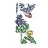
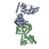
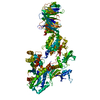
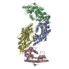
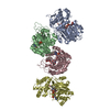
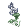
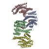
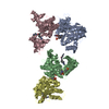
| モデル #1248 | 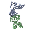 タイプ: atomic / ソフトウェア: PDB / ダミー原子の半径: 1.90 A / カイ2乗値: 0.341  Omokage検索でこの集合体の類似形状データを探す (詳細) Omokage検索でこの集合体の類似形状データを探す (詳細) |
|---|
- 試料
試料
 試料 試料 | 名称: Phosphoenolpyruvate-protein phosphotransferase / 試料濃度: 5 mg/ml |
|---|---|
| バッファ | 名称: 20mM TRIS buffer, 100 mM NaCl, 10 mM DTT, 4 mM MgCl2, 1 mM EDTA pH: 7.4 コメント: 1 tablet of protease inhibitor cocktail (SigmaFAST S8830) was added |
| 要素 #658 | 名称: Enzyme I / タイプ: protein / 記述: Phosphoenolpyruvate-protein phosphotransferase / 分子量: 63.561 / 分子数: 2 / 由来: Escherichia coli / 参照: UniProt: A0A037YGN3 配列: MISGILASPG IAFGKALLLK EDEIVIDRKK ISADQVDQEV ERFLSGRAKA SAQLETIKTK AGETFGEEKE AIFEGHIMLL EDEELEQEII ALIKDKHMTA DAAAHEVIEG QASALEELDD EYLKERAADV RDIGKRLLRN ILGLKIIDLS AIQDEVILVA ADLTPSETAQ ...配列: MISGILASPG IAFGKALLLK EDEIVIDRKK ISADQVDQEV ERFLSGRAKA SAQLETIKTK AGETFGEEKE AIFEGHIMLL EDEELEQEII ALIKDKHMTA DAAAHEVIEG QASALEELDD EYLKERAADV RDIGKRLLRN ILGLKIIDLS AIQDEVILVA ADLTPSETAQ LNLKKVLGFI TDAGGRTSHT SIMARSLELP AIVGTGSVTS QVKNDDYLIL DAVNNQVYVN PTNEVIDKMR AVQEQVASEK AELAKLKDLP AITLDGHQVE VCANIGTVRD VEGAERNGAE GVGLYRTEFL FMDRDALPTE EEQFAAYKAV AEACGSQAVI VRTMDIGGDK ELPYMNFPKE ENPFLGWRAI RIAMDRREIL RDQLRAILRA SAFGKLRIMF PMIISVEEVR ALRKEIEIYK QELRDEGKAF DESIEIGVMV ETPAAATIAR HLAKEVDFFS IGTNDLTQYT LAVDRGNDMI SHLYQPMSPS VLNLIKQVID ASHAEGKWTG MCGELAGDER ATLLLLGMGL DEFSMSAISI PRIKKIIRNT NFEDAKVLAE QALAQPTTDE LMTLVNKFIE EKTIC |
-実験情報
| ビーム | 設備名称: Advanced Photon Source (APS) 12ID-C / 地域: Argonne, IL / 国: USA  / 線源: X-ray synchrotron / 波長: 0.06199 Å / スペクトロメータ・検出器間距離: 4 mm / 線源: X-ray synchrotron / 波長: 0.06199 Å / スペクトロメータ・検出器間距離: 4 mm | ||||||||||||||||||||||||||||||
|---|---|---|---|---|---|---|---|---|---|---|---|---|---|---|---|---|---|---|---|---|---|---|---|---|---|---|---|---|---|---|---|
| 検出器 | 名称: Gold CCD | ||||||||||||||||||||||||||||||
| スキャン | 測定日: 2010年8月23日 / セル温度: 25 °C / 照射時間: 0.25 sec. / フレーム数: 20 / 単位: 1/A /
| ||||||||||||||||||||||||||||||
| 距離分布関数 P(R) |
| ||||||||||||||||||||||||||||||
| 結果 |
|
 ムービー
ムービー コントローラー


 SASDCN3
