Journal: Eur Biophys J / Year: 2017 Title: Analysis of self-assembly of S-layer protein slp-B53 from Lysinibacillus sphaericus. Authors: Jun Liu / Sven Falke / Bjoern Drobot / Dominik Oberthuer / Alexey Kikhney / Tobias Guenther / Karim Fahmy / Dmitri Svergun / Christian Betzel / Johannes Raff / Abstract: The formation of stable and functional surface layers (S-layers) via self-assembly of surface-layer proteins on the cell surface is a dynamic and complex process. S-layers facilitate a number of ...The formation of stable and functional surface layers (S-layers) via self-assembly of surface-layer proteins on the cell surface is a dynamic and complex process. S-layers facilitate a number of important biological functions, e.g., providing protection and mediating selective exchange of molecules and thereby functioning as molecular sieves. Furthermore, S-layers selectively bind several metal ions including uranium, palladium, gold, and europium, some of them with high affinity. Most current research on surface layers focuses on investigating crystalline arrays of protein subunits in Archaea and bacteria. In this work, several complementary analytical techniques and methods have been applied to examine structure-function relationships and dynamics for assembly of S-layer protein slp-B53 from Lysinibacillus sphaericus: (1) The secondary structure of the S-layer protein was analyzed by circular dichroism spectroscopy; (2) Small-angle X-ray scattering was applied to gain insights into the three-dimensional structure in solution; (3) The interaction with bivalent cations was followed by differential scanning calorimetry; (4) The dynamics and time-dependent assembly of S-layers were followed by applying dynamic light scattering; (5) The two-dimensional structure of the paracrystalline S-layer lattice was examined by atomic force microscopy. The data obtained provide essential structural insights into the mechanism of S-layer self-assembly, particularly with respect to binding of bivalent cations, i.e., Mg and Ca. Furthermore, the results obtained highlight potential applications of S-layers in the fields of micromaterials and nanobiotechnology by providing engineered or individual symmetric thin protein layers, e.g., for protective, antimicrobial, or otherwise functionalized surfaces.
Contact author
Al Kikhney (EMBL-Hamburg, European Molecular Biology Laboratory (EMBL) - Hamburg outstation, Notkestraße 85, Geb. 25A, 22607 Hamburg, Deutschland, Germany)
Instrument name: PETRA III P12 / City: Hamburg / 国: Germany / Type of source: X-ray synchrotron / Wavelength: 0.12 Å / Dist. spec. to detc.: 3.1 mm
Detector
Name: Pilatus 2M
Scan
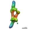
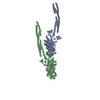
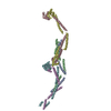
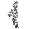
Title: slp-B53 with Mg / Measurement date: Jun 2, 2015 / Storage temperature: 20 °C / Cell temperature: 10 °C / Exposure time: 0.05 sec. / Number of frames: 20 / Unit: 1/nm /
Min
Max
Q
0.027
4.8012
Distance distribution function P(R)
Sofotware P(R): GNOM 5.0 / Number of points: 411 /
Min
Max
Q
0.0903604
1.17362
P(R) point
1
411
R
0
29
Result
Type of curve: extrapolated / Comments: slp-B53 with Mg /
Experimental
Porod
MW
178 kDa
373 kDa
Volume
-
596.6 nm3
Guinier
P(R)
Guinier error
Forward scattering, I0
8079.61
-
-
Radius of gyration, Rg
6.55 nm
7.275 nm
0.62
Min
Max
D
-
29
Guinier point
25
66
+
About Yorodumi
-
News
-
Feb 9, 2022. New format data for meta-information of EMDB entries
New format data for meta-information of EMDB entries
Version 3 of the EMDB header file is now the official format.
The previous official version 1.9 will be removed from the archive.
In the structure databanks used in Yorodumi, some data are registered as the other names, "COVID-19 virus" and "2019-nCoV". Here are the details of the virus and the list of structure data.
Jan 31, 2019. EMDB accession codes are about to change! (news from PDBe EMDB page)
EMDB accession codes are about to change! (news from PDBe EMDB page)
The allocation of 4 digits for EMDB accession codes will soon come to an end. Whilst these codes will remain in use, new EMDB accession codes will include an additional digit and will expand incrementally as the available range of codes is exhausted. The current 4-digit format prefixed with “EMD-” (i.e. EMD-XXXX) will advance to a 5-digit format (i.e. EMD-XXXXX), and so on. It is currently estimated that the 4-digit codes will be depleted around Spring 2019, at which point the 5-digit format will come into force.
The EM Navigator/Yorodumi systems omit the EMD- prefix.
Related info.:Q: What is EMD? / ID/Accession-code notation in Yorodumi/EM Navigator
Yorodumi is a browser for structure data from EMDB, PDB, SASBDB, etc.
This page is also the successor to EM Navigator detail page, and also detail information page/front-end page for Omokage search.
The word "yorodu" (or yorozu) is an old Japanese word meaning "ten thousand". "mi" (miru) is to see.
Related info.:EMDB / PDB / SASBDB / Comparison of 3 databanks / Yorodumi Search / Aug 31, 2016. New EM Navigator & Yorodumi / Yorodumi Papers / Jmol/JSmol / Function and homology information / Changes in new EM Navigator and Yorodumi
 Movie
Movie Controller

 Open data
 Basic information
 Sample
 Function and homology information
 Lysinibacillus sphaericus (bacteria)
 Citation

 Contact author
 Structure visualization
 Downloads & links
 SASDA69


 Search similar-shape structures of this assembly by Omokage search (details)