 Movie
Movie Controller


+ Open data

- Basic information
| Entry | Database: PDB / ID: 9z4q | ||||||||||||||||||||||||
|---|---|---|---|---|---|---|---|---|---|---|---|---|---|---|---|---|---|---|---|---|---|---|---|---|---|
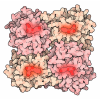
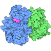
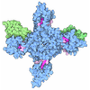
| Title | Cryo-EM structure of human nonmuscle myosin-2B, Class 3 | ||||||||||||||||||||||||
 Components Components |
| ||||||||||||||||||||||||
 Keywords Keywords | MOTOR PROTEIN / myosin / ATPase | ||||||||||||||||||||||||
| Function / homology |  Function and homology information Function and homology informationmyosin II filament / postsynaptic actin cytoskeleton organization / unconventional myosin complex / actin filament-based movement / actomyosin / myosin filament / RHO GTPases Activate ROCKs / actomyosin structure organization / RHO GTPases activate CIT / postsynaptic actin cytoskeleton ...myosin II filament / postsynaptic actin cytoskeleton organization / unconventional myosin complex / actin filament-based movement / actomyosin / myosin filament / RHO GTPases Activate ROCKs / actomyosin structure organization / RHO GTPases activate CIT / postsynaptic actin cytoskeleton / Sema4D induced cell migration and growth-cone collapse / muscle filament sliding / myosin complex / myosin II complex / skeletal muscle tissue development / EPHA-mediated growth cone collapse / structural constituent of muscle / microfilament motor activity / myosin heavy chain binding / cleavage furrow / mitotic cytokinesis / RHO GTPases activate PAKs / myofibril / cytoskeletal motor activity / brush border / Smooth Muscle Contraction / RHO GTPases activate PKNs / stress fiber / muscle contraction / positive regulation of protein secretion / neuromuscular junction / ADP binding / RNA stem-loop binding / mRNA 5'-UTR binding / spindle / Z disc / cytoplasmic side of plasma membrane / regulation of cell shape / actin filament binding / lamellipodium / growth cone / virus receptor activity / actin binding / midbody / cell cortex / vesicle / dendritic spine / calmodulin binding / cell adhesion / neuronal cell body / calcium ion binding / symbiont entry into host cell / glutamatergic synapse / cell surface / extracellular exosome / ATP binding / membrane / nucleus / cytosol / cytoplasm Similarity search - Function | ||||||||||||||||||||||||
| Biological species |  Homo sapiens (human) Homo sapiens (human) | ||||||||||||||||||||||||
| Method | ELECTRON MICROSCOPY / single particle reconstruction / cryo EM / Resolution: 4.1 Å | ||||||||||||||||||||||||
 Authors Authors | Heissler, S.M. / Chinthalapudi, K. | ||||||||||||||||||||||||
| Funding support |  United States, 1items United States, 1items
| ||||||||||||||||||||||||
 Citation Citation | Journal: Nat Commun / Year: 2026 Title: Structural basis of nonmuscle myosin-2 autoinhibition mechanisms. Authors: Sarah M Heissler / Giovanna Grandinetti / James R Sellers / Krishna Chinthalapudi / Abstract: Nonmuscle myosin-2 (NM2) is a fundamental actin-based mechanochemical ATPase that regulates cellular architecture, migration, adhesion, and force generation across diverse biological contexts. NM2 ...Nonmuscle myosin-2 (NM2) is a fundamental actin-based mechanochemical ATPase that regulates cellular architecture, migration, adhesion, and force generation across diverse biological contexts. NM2 function is tightly regulated by a structural transition between an autoinhibited monomeric (10S) conformation in which ATPase activity, actin binding, and filament assembly are coordinately suppressed and an enzymatically active, filamentous conformation. The autoinhibited conformation is critical for the spatial and temporal control of contractility in nonmuscle cells, yet structural insights into the 10S conformation remain largely elusive. Here, we report a ~53-nm elongated full-length structure of NM2B in the 10S conformation and four distinct cryo-EM structures representing the conformational landscape within the human NM2B 10S state. These structures reveal a tri-segmented tail fold that sequesters interfaces essential for actin binding and filament assembly. The asymmetric arrangement of myosin heavy and light chains provides a mechanistic foundation for understanding how regulatory post-translational modifications and disease-associated mutations shift NM2 conformational equilibria and may enable the development of structure-based interventions for cytoskeletal diseases including hearing loss, neurodegeneration, and cancer. | ||||||||||||||||||||||||
| History |
|
- Structure visualization
| Structure viewer | Molecule: MolmilJmol/JSmol |
|---|
- Downloads & links
-Download
| PDBx/mmCIF format | 9z4q.cif.gz | 683.6 KB | Display | PDBx/mmCIF format |
|---|---|---|---|---|
| PDB format | pdb9z4q.ent.gz | 543.7 KB | Display | PDB format |
| PDBx/mmJSON format | 9z4q.json.gz | Tree view | PDBx/mmJSON format | |
| Others |  Other downloads Other downloads |
-Validation report
| Arichive directory | https://data.pdbj.org/pub/pdb/validation_reports/z4/9z4qftp://data.pdbj.org/pub/pdb/validation_reports/z4/9z4q | HTTPS FTP |
|---|
-Related structure data
| Related structure data |  73811MC  9z3wC  9z3zC  9z40C C: citing same article ( M: map data used to model this data |
|---|---|
| Similar structure data |


-Links
 PDBj
PDBj










- Assembly
| Deposited unit | 
|
|---|---|
| 1 |
|
-Components
-Protein , 3 types, 6 molecules ABCFDE
| #1: Protein | Mass: 229361.547 Da / Num. of mol.: 2 Source method: isolated from a genetically manipulated source Source: (gene. exp.) Homo sapiens (human) / Gene: MYH10 / Production host:   Spodoptera frugiperda (fall armyworm) / References: UniProt: P35580 Spodoptera frugiperda (fall armyworm) / References: UniProt: P35580#2: Protein | Mass: 16948.031 Da / Num. of mol.: 2 Source method: isolated from a genetically manipulated source Details: ELC, MYL6 / Source: (gene. exp.) Homo sapiens (human) / Gene: MYL6 / Production host: Spodoptera frugiperda (fall armyworm) / References: UniProt: P60660#3: Protein | Mass: 19804.160 Da / Num. of mol.: 2 Source method: isolated from a genetically manipulated source Details: The first methionine residue is cleaved after the expression. So the first residue number is serine-1. Source: (gene. exp.) Homo sapiens (human) / Gene: MYL12B, MRLC2, MYLC2B / Production host: Spodoptera frugiperda (fall armyworm) / References: UniProt: O14950 |
|---|
-Non-polymers , 3 types, 6 molecules 

| #4: Chemical |  Mass: 24.305 Da / Num. of mol.: 2 / Source method: obtained synthetically / Formula: Mg Mass: 24.305 Da / Num. of mol.: 2 / Source method: obtained synthetically / Formula: Mg#5: Chemical | 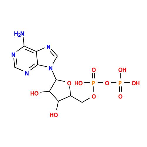 Mass: 427.201 Da / Num. of mol.: 2 / Source method: obtained synthetically / Formula: C10H15N5O10P2 / Comment: ADP, energy-carrying molecule*YM Mass: 427.201 Da / Num. of mol.: 2 / Source method: obtained synthetically / Formula: C10H15N5O10P2 / Comment: ADP, energy-carrying molecule*YM#6: Chemical | 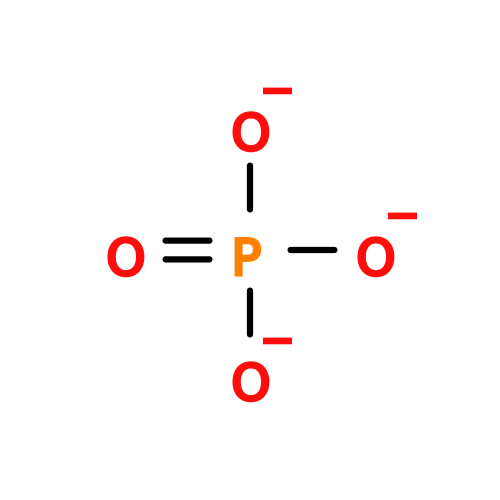 Mass: 94.971 Da / Num. of mol.: 2 / Source method: obtained synthetically / Formula: PO4 Mass: 94.971 Da / Num. of mol.: 2 / Source method: obtained synthetically / Formula: PO4 |
|---|
-Details
| Has ligand of interest | N |
|---|---|
| Has protein modification | N |
-Experimental details
-Experiment
| Experiment | Method: ELECTRON MICROSCOPY |
|---|---|
| EM experiment | Aggregation state: PARTICLE / 3D reconstruction method: single particle reconstruction |
- Sample preparation
| Component | Name: Non-muscle myosin-2B / Type: COMPLEX / Entity ID: #1-#3 / Source: RECOMBINANT |
|---|---|
| Molecular weight | Experimental value: NO |
| Source (natural) | Organism: Homo sapiens (human) |
| Source (recombinant) | Organism: Spodoptera frugiperda (fall armyworm) |
| Buffer solution | pH: 7.4 |
| Specimen | Embedding applied: NO / Shadowing applied: NO / Staining applied: NO / Vitrification applied: YES |
| Specimen support | Grid material: GOLD / Grid mesh size: 300 divisions/in. / Grid type: C-flat-1.2/1.3 |
| Vitrification | Instrument: LEICA EM GP / Cryogen name: ETHANE / Humidity: 95 % |
- Electron microscopy imaging
| Experimental equipment |  Model: Titan Krios / Image courtesy: FEI Company |
|---|---|
| Microscopy | Model: TFS KRIOS |
| Electron gun | Electron source:  FIELD EMISSION GUN / Accelerating voltage: 300 kV / Illumination mode: FLOOD BEAM FIELD EMISSION GUN / Accelerating voltage: 300 kV / Illumination mode: FLOOD BEAM |
| Electron lens | Mode: BRIGHT FIELD / Nominal defocus max: 3500 nm / Nominal defocus min: 1000 nm |
| Image recording | Electron dose: 72 e/Å2 / Detector mode: SUPER-RESOLUTION / Film or detector model: GATAN K2 QUANTUM (4k x 4k) |
- Processing
| EM software |
| |||||||||||||||||||||
|---|---|---|---|---|---|---|---|---|---|---|---|---|---|---|---|---|---|---|---|---|---|---|
| CTF correction | Type: PHASE FLIPPING AND AMPLITUDE CORRECTION | |||||||||||||||||||||
| Symmetry | Point symmetry: C1 (asymmetric) | |||||||||||||||||||||
| 3D reconstruction | Resolution: 4.1 Å / Resolution method: FSC 0.143 CUT-OFF / Num. of particles: 123125 / Symmetry type: POINT | |||||||||||||||||||||
| Atomic model building | Protocol: FLEXIBLE FIT / Space: REAL |
