[English] 日本語
Yorodumi
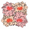
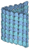
- EMDB-73815: Cryo-EM map of tail region of NM2B 10S state - class 1 -

+
Open data


ID or keywords:

Loading...

-
Basic information

Entry
Database: EMDB / ID: EMD-73815
TitleCryo-EM map of tail region of NM2B 10S state - class 1
Map dataTail region map of the 10S structure.
Sample
  • Complex: Non-muscle myosin-2B
    • Protein or peptide: MYH10 tail region
Keywordsmyosin / ATPase / MOTOR PROTEIN
Function / homology
Function and homology information


myosin II filament / postsynaptic actin cytoskeleton organization / actin filament-based movement / actomyosin / myosin filament / RHO GTPases Activate ROCKs / postsynaptic actin cytoskeleton / actomyosin structure organization / RHO GTPases activate CIT / Sema4D induced cell migration and growth-cone collapse ...myosin II filament / postsynaptic actin cytoskeleton organization / actin filament-based movement / actomyosin / myosin filament / RHO GTPases Activate ROCKs / postsynaptic actin cytoskeleton / actomyosin structure organization / RHO GTPases activate CIT / Sema4D induced cell migration and growth-cone collapse / myosin complex / myosin II complex / EPHA-mediated growth cone collapse / microfilament motor activity / cleavage furrow / mitotic cytokinesis / RHO GTPases activate PAKs / brush border / RHO GTPases activate PKNs / stress fiber / positive regulation of protein secretion / neuromuscular junction / ADP binding / RNA stem-loop binding / spindle / mRNA 5'-UTR binding / cytoplasmic side of plasma membrane / actin filament binding / regulation of cell shape / lamellipodium / growth cone / virus receptor activity / actin binding / midbody / cell cortex / dendritic spine / calmodulin binding / cell adhesion / neuronal cell body / symbiont entry into host cell / glutamatergic synapse / cell surface / extracellular exosome / ATP binding / nucleus / cytoplasm / cytosol
Similarity search - Function
IQ calmodulin-binding motif / Myosin tail / Myosin tail / Myosin N-terminal SH3-like domain / Myosin S1 fragment, N-terminal / Myosin, N-terminal, SH3-like / Myosin N-terminal SH3-like domain profile. / Short calmodulin-binding motif containing conserved Ile and Gln residues. / IQ motif, EF-hand binding site / Myosin motor domain profile. ...IQ calmodulin-binding motif / Myosin tail / Myosin tail / Myosin N-terminal SH3-like domain / Myosin S1 fragment, N-terminal / Myosin, N-terminal, SH3-like / Myosin N-terminal SH3-like domain profile. / Short calmodulin-binding motif containing conserved Ile and Gln residues. / IQ motif, EF-hand binding site / Myosin motor domain profile. / Myosin head, motor domain / Myosin head (motor domain) / Myosin. Large ATPases. / IQ motif profile. / Kinesin motor domain superfamily / P-loop containing nucleoside triphosphate hydrolase
Similarity search - Domain/homology
Biological speciesHomo sapiens (human)
Methodsingle particle reconstruction / cryo EM / Resolution: 6.64 Å
AuthorsHeissler SM / Chinthalapudi K
Funding support United States, 1 items
OrganizationGrant numberCountry
National Institutes of Health/National Institute of General Medical Sciences (NIH/NIGMS)R01GM143539 United States
CitationJournal: Nat Commun / Year: 2026
Title: Structural basis of nonmuscle myosin-2 autoinhibition mechanisms.
Authors: Sarah M Heissler / Giovanna Grandinetti / James R Sellers / Krishna Chinthalapudi /
Abstract: Nonmuscle myosin-2 (NM2) is a fundamental actin-based mechanochemical ATPase that regulates cellular architecture, migration, adhesion, and force generation across diverse biological contexts. NM2 ...Nonmuscle myosin-2 (NM2) is a fundamental actin-based mechanochemical ATPase that regulates cellular architecture, migration, adhesion, and force generation across diverse biological contexts. NM2 function is tightly regulated by a structural transition between an autoinhibited monomeric (10S) conformation in which ATPase activity, actin binding, and filament assembly are coordinately suppressed and an enzymatically active, filamentous conformation. The autoinhibited conformation is critical for the spatial and temporal control of contractility in nonmuscle cells, yet structural insights into the 10S conformation remain largely elusive. Here, we report a ~53-nm elongated full-length structure of NM2B in the 10S conformation and four distinct cryo-EM structures representing the conformational landscape within the human NM2B 10S state. These structures reveal a tri-segmented tail fold that sequesters interfaces essential for actin binding and filament assembly. The asymmetric arrangement of myosin heavy and light chains provides a mechanistic foundation for understanding how regulatory post-translational modifications and disease-associated mutations shift NM2 conformational equilibria and may enable the development of structure-based interventions for cytoskeletal diseases including hearing loss, neurodegeneration, and cancer.
History
DepositionNov 11, 2025-
Header (metadata) releaseJul 8, 2026-
Map releaseJul 8, 2026-
UpdateJul 8, 2026-
Current statusJul 8, 2026Processing site: RCSB / Status: Released

-
Structure visualization

Supplemental images

Downloads & links

-
Map

FileReleased
AnnotationTail region map of the 10S structure.
Projections & slices

Image control

Size
Brightness
Contrast
Others
AxesZ (Sec.)Y (Row.)X (Col.)
1.06 Å/pix.
x 512 pix.
= 542.72 Å
1.06 Å/pix.
x 512 pix.
= 542.72 Å
1.06 Å/pix.
x 512 pix.
= 542.72 Å

Surface

Projections

Slices (1/3)

Slices (1/2)

Slices (2/3)

Images are generated by Spider.

Voxel sizeX=Y=Z: 1.06 Å
Density
Contour LevelBy AUTHOR: 0.135
Minimum - Maximum-0.15254611 - 0.78028744
Average (Standard dev.)0.0005502675 (±0.018339746)
SymmetrySpace group: 1
Details

EMDB XML:

Map geometry
Axis orderXYZ
Origin000
Dimensions512512512
Spacing512512512
CellA=B=C: 542.72 Å
α=β=γ: 90.0 °

-
Supplemental data

-
Half map: #1

Fileemd_73815_half_map_1.map
Projections & Slices
AxesZYX

Projections

Slices (1/2)
Density Histograms

-
Half map: #2

Fileemd_73815_half_map_2.map
Projections & Slices
AxesZYX

Projections

Slices (1/2)
Density Histograms

-
Sample components

-
Entire : Non-muscle myosin-2B

EntireName: Non-muscle myosin-2B
Components
  • Complex: Non-muscle myosin-2B
    • Protein or peptide: MYH10 tail region

-
Supramolecule #1: Non-muscle myosin-2B

SupramoleculeName: Non-muscle myosin-2B / type: complex / ID: 1 / Parent: 0 / Macromolecule list: all
Source (natural)Organism: Homo sapiens (human)

-
Macromolecule #1: MYH10 tail region

MacromoleculeName: MYH10 tail region / type: protein_or_peptide / ID: 1 / Details: coiled coil tail region. / Enantiomer: LEVO
Source (natural)Organism: Homo sapiens (human)
Recombinant expressionOrganism: Spodoptera frugiperda (fall armyworm)
SequenceString: MAQRTGLEDP ERYLFVDRAV IYNPATQADW TAKKLVWIPS ERHGFEAASI KEERGDEVMV ELAENGKKAM VNKDDIQKMN PPKFSKVEDM AELTCLNEAS VLHNLKDRYY SGLIYTYSGL FCVVINPYKN LPIYSENIIE MYRGKKRHEM PPHIYAISES AYRCMLQDRE ...String:
MAQRTGLEDP ERYLFVDRAV IYNPATQADW TAKKLVWIPS ERHGFEAASI KEERGDEVMV ELAENGKKAM VNKDDIQKMN PPKFSKVEDM AELTCLNEAS VLHNLKDRYY SGLIYTYSGL FCVVINPYKN LPIYSENIIE MYRGKKRHEM PPHIYAISES AYRCMLQDRE DQSILCTGES GAGKTENTKK VIQYLAHVAS SHKGRKDHNI PGELERQLLQ ANPILESFGN AKTVKNDNSS RFGKFIRINF DVTGYIVGAN IETYLLEKSR AVRQAKDERT FHIFYQLLSG AGEHLKSDLL LEGFNNYRFL SNGYIPIPGQ QDKDNFQETM EAMHIMGFSH EEILSMLKVV SSVLQFGNIS FKKERNTDQA SMPENTVAQK LCHLLGMNVM EFTRAILTPR IKVGRDYVQK AQTKEQADFA VEALAKATYE RLFRWLVHRI NKALDRTKRQ GASFIGILDI AGFEIFELNS FEQLCINYTN EKLQQLFNHT MFILEQEEYQ REGIEWNFID FGLDLQPCID LIERPANPPG VLALLDEECW FPKATDKTFV EKLVQEQGSH SKFQKPRQLK DKADFCIIHY AGKVDYKADE WLMKNMDPLN DNVATLLHQS SDRFVAELWK DVDRIVGLDQ VTGMTETAFG SAYKTKKGMF RTVGQLYKES LTKLMATLRN TNPNFVRCII PNHEKRAGKL DPHLVLDQLR CNGVLEGIRI CRQGFPNRIV FQEFRQRYEI LTPNAIPKGF MDGKQACERM IRALELDPNL YRIGQSKIFF RAGVLAHLEE ERDLKITDII IFFQAVCRGY LARKAFAKKQ QQLSALKVLQ RNCAAYLKLR HWQWWRVFTK VKPLLQVTRQ EEELQAKDEE LLKVKEKQTK VEGELEEMER KHQQLLEEKN ILAEQLQAET ELFAEAEEMR ARLAAKKQEL EEILHDLESR VEEEEERNQI LQNEKKKMQA HIQDLEEQLD EEEGARQKLQ LEKVTAEAKI KKMEEEILLL EDQNSKFIKE KKLMEDRIAE CSSQLAEEEE KAKNLAKIRN KQEVMISDLE ERLKKEEKTR QELEKAKRKL DGETTDLQDQ IAELQAQIDE LKLQLAKKEE ELQGALARGD DETLHKNNAL KVVRELQAQI AELQEDFESE KASRNKAEKQ KRDLSEELEA LKTELEDTLD TTAAQQELRT KREQEVAELK KALEEETKNH EAQIQDMRQR HATALEELSE QLEQAKRFKA NLEKNKQGLE TDNKELACEV KVLQQVKAES EHKRKKLDAQ VQELHAKVSE GDRLRVELAE KASKLQNELD NVSTLLEEAE KKGIKFAKDA ASLESQLQDT QELLQEETRQ KLNLSSRIRQ LEEEKNSLQE QQEEEEEARK NLEKQVLALQ SQLADTKKKV DDDLGTIESL EEAKKKLLKD AEALSQRLEE KALAYDKLEK TKNRLQQELD DLTVDLDHQR QVASNLEKKQ KKFDQLLAEE KSISARYAEE RDRAEAEARE KETKALSLAR ALEEALEAKE EFERQNKQLR ADMEDLMSSK DDVGKNVHEL EKSKRALEQQ VEEMRTQLEE LEDELQATED AKLRLEVNMQ AMKAQFERDL QTRDEQNEEK KRLLIKQVRE LEAELEDERK QRALAVASKK KMEIDLKDLE AQIEAANKAR DEVIKQLRKL QAQMKDYQRE LEEARASRDE IFAQSKESEK KLKSLEAEIL QLQEELASSE RARRHAEQER DELADEITNS ASGKSALLDE KRRLEARIAQ LEEELEEEQS NMELLNDRFR KTTLQVDTLN AELAAERSAA QKSDNARQQL ERQNKELKAK LQELEGAVKS KFKATISALE AKIGQLEEQL EQEAKERAAA NKLVRRTEKK LKEIFMQVED ERRHADQYKE QMEKANARMK QLKRQLEEAE EEATRANASR RKLQRELDDA TEANEGLSRE VSTLKNRLRR GGPISFSSSR SGRRQLHLEG ASLELSDDDT ESKTSDVNET QPPQSE

UniProtKB: Myosin-10

-
Experimental details

-
Structure determination

Methodcryo EM
Processingsingle particle reconstruction
Aggregation stateparticle

-
Sample preparation

BufferpH: 7.4
GridModel: C-flat-1.2/1.3 / Material: GOLD / Mesh: 300
VitrificationCryogen name: ETHANE

-
Electron microscopy

MicroscopeTFS KRIOS
Image recordingFilm or detector model: GATAN K2 QUANTUM (4k x 4k) / Detector mode: SUPER-RESOLUTION / Average electron dose: 72.0 e/Å2
Electron beamAcceleration voltage: 300 kV / Electron source: FIELD EMISSION GUN
Electron opticsIllumination mode: FLOOD BEAM / Imaging mode: BRIGHT FIELD / Nominal defocus max: 3.5 µm / Nominal defocus min: 1.0 µm
Experimental equipment
Model: Titan Krios / Image courtesy: FEI Company

+
Image processing

CTF correctionSoftware - Name: cryoSPARC / Type: PHASE FLIPPING AND AMPLITUDE CORRECTION
Startup modelType of model: PDB ENTRY
PDB model - PDB ID:
Final reconstructionResolution.type: BY AUTHOR / Resolution: 6.64 Å / Resolution method: FSC 0.143 CUT-OFF / Software - Name: cryoSPARC / Number images used: 25065
Initial angle assignmentType: MAXIMUM LIKELIHOOD / Software - Name: cryoSPARC
Final angle assignmentType: MAXIMUM LIKELIHOOD / Software - Name: cryoSPARC
FSC plot (resolution estimation)

-
Atomic model buiding 1

RefinementSpace: REAL / Protocol: FLEXIBLE FIT

+
About Yorodumi

-
News

-
Feb 9, 2022. New format data for meta-information of EMDB entries

New format data for meta-information of EMDB entries

  • Version 3 of the EMDB header file is now the official format.
  • The previous official version 1.9 will be removed from the archive.

Related info.:EMDB header

External links:wwPDB to switch to version 3 of the EMDB data model

-
Aug 12, 2020. Covid-19 info

Covid-19 info

URL: https://pdbj.org/emnavi/covid19.php

New page: Covid-19 featured information page in EM Navigator.

Related info.:Covid-19 info / Mar 5, 2020. Novel coronavirus structure data

+
Mar 5, 2020. Novel coronavirus structure data

Novel coronavirus structure data

Related info.:Yorodumi Speices / Aug 12, 2020. Covid-19 info

External links:COVID-19 featured content - PDBj / Molecule of the Month (242):Coronavirus Proteases

+
Jan 31, 2019. EMDB accession codes are about to change! (news from PDBe EMDB page)

EMDB accession codes are about to change! (news from PDBe EMDB page)

  • The allocation of 4 digits for EMDB accession codes will soon come to an end. Whilst these codes will remain in use, new EMDB accession codes will include an additional digit and will expand incrementally as the available range of codes is exhausted. The current 4-digit format prefixed with “EMD-” (i.e. EMD-XXXX) will advance to a 5-digit format (i.e. EMD-XXXXX), and so on. It is currently estimated that the 4-digit codes will be depleted around Spring 2019, at which point the 5-digit format will come into force.
  • The EM Navigator/Yorodumi systems omit the EMD- prefix.

Related info.:Q: What is EMD? / ID/Accession-code notation in Yorodumi/EM Navigator

External links:EMDB Accession Codes are Changing Soon! / Contact to PDBj

+
Jul 12, 2017. Major update of PDB

Major update of PDB

  • wwPDB released updated PDB data conforming to the new PDBx/mmCIF dictionary.
  • This is a major update changing the version number from 4 to 5, and with Remediation, in which all the entries are updated.
  • In this update, many items about electron microscopy experimental information are reorganized (e.g. em_software).
  • Now, EM Navigator and Yorodumi are based on the updated data.

External links:wwPDB Remediation / Enriched Model Files Conforming to OneDep Data Standards Now Available in the PDB FTP Archive

-
Yorodumi

Thousand views of thousand structures

  • Yorodumi is a browser for structure data from EMDB, PDB, SASBDB, etc.
  • This page is also the successor to EM Navigator detail page, and also detail information page/front-end page for Omokage search.
  • The word "yorodu" (or yorozu) is an old Japanese word meaning "ten thousand". "mi" (miru) is to see.

Related info.:EMDB / PDB / SASBDB / Comparison of 3 databanks / Yorodumi Search / Aug 31, 2016. New EM Navigator & Yorodumi / Yorodumi Papers / Jmol/JSmol / Function and homology information / Changes in new EM Navigator and Yorodumi

Read more