Movie
Movie Controller

[English] 日本語
 Yorodumi
- PDB-9yp9: Cryo-EM structure of human beta-cardiac myosin bound to mavacamte... -
+ Open data

- Basic information
| Entry | Database: PDB / ID: 9yp9 | |||||||||||||||||||||||||||
|---|---|---|---|---|---|---|---|---|---|---|---|---|---|---|---|---|---|---|---|---|---|---|---|---|---|---|---|---|
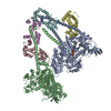
| Title | Cryo-EM structure of human beta-cardiac myosin bound to mavacamten in the interacting-heads motif and S2-FH docked state | |||||||||||||||||||||||||||
 Components Components |
| |||||||||||||||||||||||||||
 Keywords Keywords | CONTRACTILE PROTEIN / Cardiac Myosin / Interacting Heads Motif / Mavacamten / Actin Binding | |||||||||||||||||||||||||||
| Function / homology |  Function and homology information Function and homology informationregulation of slow-twitch skeletal muscle fiber contraction / regulation of the force of skeletal muscle contraction / unconventional myosin complex / Striated Muscle Contraction / contractile muscle fiber / Smooth Muscle Contraction / FCERI mediated MAPK activation / protein localization to nuclear periphery / muscle myosin complex / Activation of the AP-1 family of transcription factors ...regulation of slow-twitch skeletal muscle fiber contraction / regulation of the force of skeletal muscle contraction / unconventional myosin complex / Striated Muscle Contraction / contractile muscle fiber / Smooth Muscle Contraction / FCERI mediated MAPK activation / protein localization to nuclear periphery / muscle myosin complex / Activation of the AP-1 family of transcription factors / negative regulation of ribosomal protein gene transcription by RNA polymerase II / positive regulation of cellular response to amino acid starvation / response to amino acid starvation / mediator complex binding / regulation of the force of heart contraction / transition between fast and slow fiber / myosin filament / Oxidative Stress Induced Senescence / cardiac muscle hypertrophy in response to stress / muscle filament sliding / myosin complex / adult heart development / myosin II complex / structural constituent of muscle / ventricular cardiac muscle tissue morphogenesis / microfilament motor activity / myofibril / amino acid biosynthetic process / TFIID-class transcription factor complex binding / cytoskeletal motor activity / positive regulation of RNA polymerase II transcription preinitiation complex assembly / positive regulation of transcription initiation by RNA polymerase II / skeletal muscle tissue development / ATP metabolic process / striated muscle contraction / skeletal muscle contraction / cardiac muscle contraction / stress fiber / cellular response to nutrient levels / regulation of heart rate / muscle contraction / cellular response to amino acid starvation / bioluminescence / sarcomere / generation of precursor metabolites and energy / RNA polymerase II transcription regulator complex / Z disc / actin filament binding / DNA-binding transcription activator activity, RNA polymerase II-specific / transcription regulator complex / sequence-specific DNA binding / RNA polymerase II-specific DNA-binding transcription factor binding / DNA-binding transcription factor activity, RNA polymerase II-specific / calmodulin binding / intracellular signal transduction / RNA polymerase II cis-regulatory region sequence-specific DNA binding / DNA-binding transcription factor activity / calcium ion binding / chromatin binding / negative regulation of transcription by RNA polymerase II / positive regulation of transcription by RNA polymerase II / ATP binding / identical protein binding / nucleus / cytoplasm Similarity search - Function | |||||||||||||||||||||||||||
| Biological species |  Homo sapiens (human) Homo sapiens (human)   Aequorea victoria (jellyfish) Aequorea victoria (jellyfish) | |||||||||||||||||||||||||||
| Method | ELECTRON MICROSCOPY / single particle reconstruction / cryo EM / Resolution: 3 Å | |||||||||||||||||||||||||||
 Authors Authors | Somavarapu, A.K. / Ge, J. / Yengo, C.M. / Craig, R. / Padron, R. | |||||||||||||||||||||||||||
| Funding support |  United States, 2items United States, 2items
| |||||||||||||||||||||||||||
 Citation Citation | Journal: Sci Adv / Year: 2026 Title: Cryo-EM reveals how cardiomyopathy therapeutic drugs modulate the myosin motors of the heart. Authors: Arun Kumar Somavarapu / Jinghua Ge / Christopher M Yengo / Roger Craig / Raul Padron / Abstract: Genetic mutations in myosin, the motor protein that powers the heartbeat, are linked to inherited hypertrophic and dilated cardiomyopathies. Mavacamten and omecamtiv mecarbil are therapeutic, myosin- ...Genetic mutations in myosin, the motor protein that powers the heartbeat, are linked to inherited hypertrophic and dilated cardiomyopathies. Mavacamten and omecamtiv mecarbil are therapeutic, myosin-targeted drugs designed to treat these myopathies, but their mechanism of action has remained unclear. Using single-particle cryo-EM, we determined near-atomic resolution structures of wild-type, mavacamten-bound, and omecamtiv mecarbil-bound myosin molecules. Across all conditions, we observe two distinct, alternate conformations of myosin, not previously reported. We show how mavacamten stabilizes one conformation by reinforcing key electrostatic interfaces in the molecule, whereas omecamtiv mecarbil weakens these interfaces, favoring the second structure. This remodeling elucidates previously unclear allosteric mechanisms through which these drugs either inhibit or enhance myosin activity, countering the deleterious impacts of disease. These findings reveal how drugs modulate myosin structure to control cardiac contractility. | |||||||||||||||||||||||||||
| History |
|
- Structure visualization
| Structure viewer | Molecule: MolmilJmol/JSmol |
|---|
- Downloads & links
-Download
| PDBx/mmCIF format | 9yp9.cif.gz | 511.2 KB | Display | PDBx/mmCIF format |
|---|---|---|---|---|
| PDB format | pdb9yp9.ent.gz | 406.4 KB | Display | PDB format |
| PDBx/mmJSON format | 9yp9.json.gz | Tree view | PDBx/mmJSON format | |
| Others |  Other downloads Other downloads |
-Validation report
| Arichive directory | https://data.pdbj.org/pub/pdb/validation_reports/yp/9yp9ftp://data.pdbj.org/pub/pdb/validation_reports/yp/9yp9 | HTTPS FTP |
|---|
-Related structure data
| Related structure data |  73288MC  9yopC  9yp4C  9yr7C  9yrgC  9yrhC M: map data used to model this data C: citing same article ( |
|---|---|
| Similar structure data |


-Links
 PDBj
PDBj


- Assembly
| Deposited unit | 
|
|---|---|
| 1 |
|
-Components
-Protein , 3 types, 6 molecules ABCDEF
| #1: Protein | Mass: 150656.500 Da / Num. of mol.: 2 Source method: isolated from a genetically manipulated source Source: (gene. exp.) Homo sapiens (human), (gene. exp.) Aequorea victoria (jellyfish)Gene: MYH7, MYHCB, GCN4, AAS101, AAS3, ARG9, YEL009C, GFP / Production host: References: UniProt: P12883, UniProt: P03069, UniProt: P42212 #2: Protein | Mass: 20620.490 Da / Num. of mol.: 2 / Source method: isolated from a natural source / Source: (natural) #3: Protein | Mass: 18978.445 Da / Num. of mol.: 2 / Source method: isolated from a natural source / Source: (natural) |
|---|
-Non-polymers , 3 types, 6 molecules 


| #4: Chemical |  Mass: 94.971 Da / Num. of mol.: 2 / Source method: obtained synthetically / Formula: PO4 / Feature type: SUBJECT OF INVESTIGATION Mass: 94.971 Da / Num. of mol.: 2 / Source method: obtained synthetically / Formula: PO4 / Feature type: SUBJECT OF INVESTIGATION#5: Chemical |  Mass: 273.330 Da / Num. of mol.: 2 / Source method: obtained synthetically / Formula: C15H19N3O2 / Feature type: SUBJECT OF INVESTIGATION Mass: 273.330 Da / Num. of mol.: 2 / Source method: obtained synthetically / Formula: C15H19N3O2 / Feature type: SUBJECT OF INVESTIGATION#6: Chemical |  Mass: 427.201 Da / Num. of mol.: 2 / Source method: obtained synthetically / Formula: C10H15N5O10P2 / Feature type: SUBJECT OF INVESTIGATION / Comment: ADP, energy-carrying molecule*YM Mass: 427.201 Da / Num. of mol.: 2 / Source method: obtained synthetically / Formula: C10H15N5O10P2 / Feature type: SUBJECT OF INVESTIGATION / Comment: ADP, energy-carrying molecule*YM |
|---|
-Details
| Has ligand of interest | Y |
|---|---|
| Has protein modification | N |
-Experimental details
-Experiment
| Experiment | Method: ELECTRON MICROSCOPY |
|---|---|
| EM experiment | Aggregation state: PARTICLE / 3D reconstruction method: single particle reconstruction |
- Sample preparation
| Component |
| ||||||||||||||||||||||||
|---|---|---|---|---|---|---|---|---|---|---|---|---|---|---|---|---|---|---|---|---|---|---|---|---|---|
| Molecular weight | Value: 0.11652168 MDa / Experimental value: NO | ||||||||||||||||||||||||
| Source (natural) |
| ||||||||||||||||||||||||
| Source (recombinant) | Organism: | ||||||||||||||||||||||||
| Buffer solution | pH: 7.4 | ||||||||||||||||||||||||
| Specimen | Embedding applied: NO / Shadowing applied: NO / Staining applied: NO / Vitrification applied: YES | ||||||||||||||||||||||||
| Specimen support | Grid material: GOLD / Grid type: UltrAuFoil R1.2/1.3 | ||||||||||||||||||||||||
| Vitrification | Instrument: FEI VITROBOT MARK IV / Cryogen name: ETHANE |
- Electron microscopy imaging
| Experimental equipment |  Model: Titan Krios / Image courtesy: FEI Company |
|---|---|
| Microscopy | Model: TFS KRIOS |
| Electron gun | Electron source:  FIELD EMISSION GUN / Accelerating voltage: 300 kV / Illumination mode: FLOOD BEAM FIELD EMISSION GUN / Accelerating voltage: 300 kV / Illumination mode: FLOOD BEAM |
| Electron lens | Mode: BRIGHT FIELD / Nominal defocus max: 2000 nm / Nominal defocus min: 1000 nm |
| Specimen holder | Specimen holder model: FEI TITAN KRIOS AUTOGRID HOLDER |
| Image recording | Electron dose: 49.5 e/Å2 / Film or detector model: GATAN K3 (6k x 4k) |
- Processing
| EM software |
| |||||||||||||||||||||||||||||||||||||||||||||||||||||||||||||||||
|---|---|---|---|---|---|---|---|---|---|---|---|---|---|---|---|---|---|---|---|---|---|---|---|---|---|---|---|---|---|---|---|---|---|---|---|---|---|---|---|---|---|---|---|---|---|---|---|---|---|---|---|---|---|---|---|---|---|---|---|---|---|---|---|---|---|---|
| CTF correction | Type: PHASE FLIPPING AND AMPLITUDE CORRECTION | |||||||||||||||||||||||||||||||||||||||||||||||||||||||||||||||||
| 3D reconstruction | Resolution: 3 Å / Resolution method: FSC 0.143 CUT-OFF / Num. of particles: 216820 / Symmetry type: POINT | |||||||||||||||||||||||||||||||||||||||||||||||||||||||||||||||||
| Atomic model building |
| |||||||||||||||||||||||||||||||||||||||||||||||||||||||||||||||||
| Atomic model building | Source name: PDB / Type: experimental model
|