Movie
Movie Controller

[English] 日本語
 Yorodumi
- EMDB-73362: Cryo-EM structure of human beta-cardiac myosin bound to mavacamte... -
+ Open data

- Basic information
| Entry |  | |||||||||
|---|---|---|---|---|---|---|---|---|---|---|
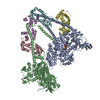
| Title | Cryo-EM structure of human beta-cardiac myosin bound to mavacamten in the interacting-heads motif and S2-FH undocked state | |||||||||
 Map data Map data | ||||||||||
 Sample Sample |
| |||||||||
 Keywords Keywords | Cardiac Myosin / Interacting Heads Motif / Mavacamten / Actin Binding / CONTRACTILE PROTEIN | |||||||||
| Function / homology |  Function and homology information Function and homology informationregulation of slow-twitch skeletal muscle fiber contraction / regulation of the force of skeletal muscle contraction / Striated Muscle Contraction / unconventional myosin complex / contractile muscle fiber / Smooth Muscle Contraction / FCERI mediated MAPK activation / protein localization to nuclear periphery / Activation of the AP-1 family of transcription factors / muscle myosin complex ...regulation of slow-twitch skeletal muscle fiber contraction / regulation of the force of skeletal muscle contraction / Striated Muscle Contraction / unconventional myosin complex / contractile muscle fiber / Smooth Muscle Contraction / FCERI mediated MAPK activation / protein localization to nuclear periphery / Activation of the AP-1 family of transcription factors / muscle myosin complex / negative regulation of ribosomal protein gene transcription by RNA polymerase II / positive regulation of cellular response to amino acid starvation / response to amino acid starvation / mediator complex binding / regulation of the force of heart contraction / transition between fast and slow fiber / myosin filament / Oxidative Stress Induced Senescence / cardiac muscle hypertrophy in response to stress / muscle filament sliding / adult heart development / myosin complex / myosin II complex / structural constituent of muscle / ventricular cardiac muscle tissue morphogenesis / microfilament motor activity / amino acid biosynthetic process / TFIID-class transcription factor complex binding / myofibril / cytoskeletal motor activity / positive regulation of RNA polymerase II transcription preinitiation complex assembly / positive regulation of transcription initiation by RNA polymerase II / skeletal muscle tissue development / ATP metabolic process / striated muscle contraction / skeletal muscle contraction / cardiac muscle contraction / stress fiber / cellular response to nutrient levels / regulation of heart rate / muscle contraction / cellular response to amino acid starvation / bioluminescence / sarcomere / generation of precursor metabolites and energy / RNA polymerase II transcription regulator complex / Z disc / actin filament binding / DNA-binding transcription activator activity, RNA polymerase II-specific / transcription regulator complex / sequence-specific DNA binding / RNA polymerase II-specific DNA-binding transcription factor binding / DNA-binding transcription factor activity, RNA polymerase II-specific / calmodulin binding / intracellular signal transduction / RNA polymerase II cis-regulatory region sequence-specific DNA binding / DNA-binding transcription factor activity / calcium ion binding / chromatin binding / negative regulation of transcription by RNA polymerase II / positive regulation of transcription by RNA polymerase II / ATP binding / identical protein binding / nucleus / cytoplasm Similarity search - Function | |||||||||
| Biological species |  Homo sapiens (human) / Homo sapiens (human) /    Aequorea victoria (jellyfish) Aequorea victoria (jellyfish) | |||||||||
| Method | single particle reconstruction / cryo EM / Resolution: 3.0 Å | |||||||||
 Authors Authors | Somavarapu AK / Ge J / Yengo CM / Craig R / Padron R | |||||||||
| Funding support |  United States, 2 items United States, 2 items
| |||||||||
 Citation Citation | Journal: Sci Adv / Year: 2026 Title: Cryo-EM reveals how cardiomyopathy therapeutic drugs modulate the myosin motors of the heart. Authors: Arun Kumar Somavarapu / Jinghua Ge / Christopher M Yengo / Roger Craig / Raul Padron / Abstract: Genetic mutations in myosin, the motor protein that powers the heartbeat, are linked to inherited hypertrophic and dilated cardiomyopathies. Mavacamten and omecamtiv mecarbil are therapeutic, myosin- ...Genetic mutations in myosin, the motor protein that powers the heartbeat, are linked to inherited hypertrophic and dilated cardiomyopathies. Mavacamten and omecamtiv mecarbil are therapeutic, myosin-targeted drugs designed to treat these myopathies, but their mechanism of action has remained unclear. Using single-particle cryo-EM, we determined near-atomic resolution structures of wild-type, mavacamten-bound, and omecamtiv mecarbil-bound myosin molecules. Across all conditions, we observe two distinct, alternate conformations of myosin, not previously reported. We show how mavacamten stabilizes one conformation by reinforcing key electrostatic interfaces in the molecule, whereas omecamtiv mecarbil weakens these interfaces, favoring the second structure. This remodeling elucidates previously unclear allosteric mechanisms through which these drugs either inhibit or enhance myosin activity, countering the deleterious impacts of disease. These findings reveal how drugs modulate myosin structure to control cardiac contractility. | |||||||||
| History |
|
- Structure visualization
| Supplemental images |
|---|

- Downloads & links
-EMDB archive
| Map data | emd_73362.map.gz | 125.4 MB | EMDB map data format | |
|---|---|---|---|---|
| Header (meta data) | emd-73362-v30.xmlemd-73362.xml | 26.8 KB 26.8 KB | Display Display | EMDB header |
| FSC (resolution estimation) | emd_73362_fsc.xml | 13.3 KB | Display | FSC data file |
| Images |  emd_73362.png emd_73362.png | 105.6 KB | ||
| Masks | emd_73362_msk_1.map | 244.1 MB | Mask map | |
| Filedesc metadata | emd-73362.cif.gz | 8.2 KB | ||
| Others | emd_73362_half_map_1.map.gzemd_73362_half_map_2.map.gz | 226.9 MB 226.9 MB | ||
| Archive directory |  http://ftp.pdbj.org/pub/emdb/structures/EMD-73362ftp://ftp.pdbj.org/pub/emdb/structures/EMD-73362 http://ftp.pdbj.org/pub/emdb/structures/EMD-73362ftp://ftp.pdbj.org/pub/emdb/structures/EMD-73362 | HTTPS FTP |
-Related structure data
| Related structure data |  9yr7MC  9yopC  9yp4C  9yp9C  9yrgC  9yrhC M: atomic model generated by this map C: citing same article ( |
|---|---|
| Similar structure data |


-Links
| EMDB pages | EMDB (EBI/PDBe) / EMDataResource |
|---|---|
| Related items in Molecule of the Month |


-Map
| File | Download / File: emd_73362.map.gz / Format: CCP4 / Size: 244.1 MB / Type: IMAGE STORED AS FLOATING POINT NUMBER (4 BYTES) | ||||||||||||||||||||||||||||||||||||
|---|---|---|---|---|---|---|---|---|---|---|---|---|---|---|---|---|---|---|---|---|---|---|---|---|---|---|---|---|---|---|---|---|---|---|---|---|---|
| Projections & slices | Image control
Images are generated by Spider. | ||||||||||||||||||||||||||||||||||||
| Voxel size | X=Y=Z: 0.83 Å | ||||||||||||||||||||||||||||||||||||
| Density |
| ||||||||||||||||||||||||||||||||||||
| Symmetry | Space group: 1 | ||||||||||||||||||||||||||||||||||||
| Details | EMDB XML:
|
 Z (Sec.)
 Y (Row.)
 X (Col.)


-Supplemental data
-Mask #1
| File | emd_73362_msk_1.map | ||||||||||||
|---|---|---|---|---|---|---|---|---|---|---|---|---|---|
| Projections & Slices |
| ||||||||||||
| Density Histograms |


-Half map: #1
| File | emd_73362_half_map_1.map | ||||||||||||
|---|---|---|---|---|---|---|---|---|---|---|---|---|---|
| Projections & Slices |
| ||||||||||||
| Density Histograms |


-Half map: #2
| File | emd_73362_half_map_2.map | ||||||||||||
|---|---|---|---|---|---|---|---|---|---|---|---|---|---|
| Projections & Slices |
| ||||||||||||
| Density Histograms |


- Sample components
-Entire : Human beta-cardiac myosin bound to mavacamten in the interacting-...
| Entire | Name: Human beta-cardiac myosin bound to mavacamten in the interacting-heads motif and S2-FH undocked state |
|---|---|
| Components |
|
-Supramolecule #1: Human beta-cardiac myosin bound to mavacamten in the interacting-...
| Supramolecule | Name: Human beta-cardiac myosin bound to mavacamten in the interacting-heads motif and S2-FH undocked state type: complex / ID: 1 / Parent: 0 / Macromolecule list: #1-#3 |
|---|---|
| Molecular weight | Theoretical: 116.52168 KDa |
-Supramolecule #2: beta-cardiac myosin
| Supramolecule | Name: beta-cardiac myosin / type: complex / ID: 2 / Parent: 1 / Macromolecule list: #1 |
|---|---|
| Source (natural) | Organism: Homo sapiens (human) |
-Supramolecule #3: Myosin light chain 1/3 and Myosin regulatory light chain 11
| Supramolecule | Name: Myosin light chain 1/3 and Myosin regulatory light chain 11 type: complex / ID: 3 / Parent: 1 / Macromolecule list: #2-#3 |
|---|---|
| Source (natural) | Organism: |
-Macromolecule #1: Myosin-7,General control transcription factor GCN4,Enhanced Green...
| Macromolecule | Name: Myosin-7,General control transcription factor GCN4,Enhanced Green fluorescent protein type: protein_or_peptide / ID: 1 / Number of copies: 2 / Enantiomer: LEVO |
|---|---|
| Source (natural) | Organism: Aequorea victoria (jellyfish) |
| Molecular weight | Theoretical: 150.6565 KDa |
| Recombinant expression | Organism: |
| Sequence | String: MGDSEMAVFG AAAPYLRKSE KERLEAQTRP FDLKKDVFVP DDKQEFVKAK IVSREGGKVT AETEYGKTVT VKEDQVMQQN PPKFDKIED MAMLTFLHEP AVLYNLKDRY GSWMIYTYSG LFCVTVNPYK WLPVYTPEVV AAYRGKKRSE APPHIFSISD N AYQYMLTD ...String: MGDSEMAVFG AAAPYLRKSE KERLEAQTRP FDLKKDVFVP DDKQEFVKAK IVSREGGKVT AETEYGKTVT VKEDQVMQQN PPKFDKIED MAMLTFLHEP AVLYNLKDRY GSWMIYTYSG LFCVTVNPYK WLPVYTPEVV AAYRGKKRSE APPHIFSISD N AYQYMLTD RENQSILITG ESGAGKTVNT KRVIQYFAVI AAIGDRSKKD QSPGKGTLED QIIQANPALE AFGNAKTVRN DN SSRFGKF IRIHFGATGK LASADIETYL LEKSRVIFQL KAERDYHIFY QILSNKKPEL LDMLLITNNP YDYAFISQGE TTV ASIDDA EELMATDNAF DVLGFTSEEK NSMYKLTGAI MHFGNMKFKL KQREEQAEPD GTEEADKSAY LMGLNSADLL KGLC HPRVK VGNEYVTKGQ NVQQVIYATG ALAKAVYERM FNWMVTRINA TLETKQPRQY FIGVLDIAGF EIFDFNSFEQ LCINF TNEK LQQFFNHHMF VLEQEEYKKE GIEWTFIDFG MDLQACIDLI EKPMGIMSIL EEECMFPKAT DMTFKAKLFD NHLGKS ANF QKPRNIKGKP EAHFSLIHYA GIVDYNIIGW LQKNKDPLNE TVVGLYQKSS LKLLSTLFAN YAGADAPIEK GKGKAKK GS SFQTVSALHR ENLNKLMTNL RSTHPHFVRC IIPNETKSPG VMDNPLVMHQ LRCNGVLEGI RICRKGFPNR ILYGDFRQ R YRILNPAAIP EGQFIDSRKG AEKLLSSLDI DHNQYKFGHT KVFFKAGLLG LLEEMRDERL SRIITRIQAQ SRGVLARME YKKLLERRDS LLVIQWNIRA FMGVKNWPWM KLYFKIKPLL KSAEREKEMA SMKEEFTRLK EALEKSEARR KELEEKMVSL LQEKNDLQL QVQAEQDNLA DAEERCDQLI KNKIQLEAKV KEMNERLEDE EEMNAELTAK KRKLEDECSE LKRDIDDLEL T LAKVEKEK HATENKVKNL TEEMAGLDEI IAKLTKEKKA LQEAHQQALD DLQAEEDKMK QLEDKVEELL SKNYHLENEV AR LKKLVGE RGSGKLGVSK GEELFTGVVP ILVELDGDVN GHKFSVSGEG EGDATYGKLT LKFICTTGKL PVPWPTLVTT LTY GVQCFS RYPDHMKQHD FFKSAMPEGY VQERTIFFKD DGNYKTRAEV KFEGDTLVNR IELKGIDFKE DGNILGHKLE YNYN SHNVY IMADKQKNGI KVNFKIRHNI EDGSVQLADH YQQNTPIGDG PVLLPDNHYL STQSALSKDP NEKRDHMVLL EFVTA AGIT LGMDELYKGL NDIFEAQKIE WHEDYKDDDD K UniProtKB: Myosin-7, General control transcription factor GCN4, Green fluorescent protein |
-Macromolecule #2: Myosin light chain 1/3, skeletal muscle isoform
| Macromolecule | Name: Myosin light chain 1/3, skeletal muscle isoform / type: protein_or_peptide / ID: 2 / Number of copies: 2 / Enantiomer: LEVO |
|---|---|
| Source (natural) | Organism: |
| Molecular weight | Theoretical: 20.62049 KDa |
| Sequence | String: MAPKKDVKKP AAAPAPAPAP APAPAKPKEE KIDLSAIKIE FSKEQQEDFK EAFLLFDRTG ECKITLSQVG DVLRALGTNP TNAEVKKVL GNPSNEEMNA KKIEFEQFLP MMQAISNNKD QGGYEDFVEG LRVFDKEGNG TVMGAELRHV LATLGEKMKE E EVEALLAG QEDSNGCINY EAFVKHIMSV UniProtKB: Myosin light chain 1/3, skeletal muscle isoform |
-Macromolecule #3: Myosin regulatory light chain 11
| Macromolecule | Name: Myosin regulatory light chain 11 / type: protein_or_peptide / ID: 3 / Number of copies: 2 / Enantiomer: LEVO |
|---|---|
| Source (natural) | Organism: |
| Molecular weight | Theoretical: 18.978445 KDa |
| Sequence | String: MAPKKAKRRA GAEGSSNVFS MFDQTQIQEF KEAFTVIDQN RDGIIDKEDL RDTFAAMGRL NVKNEELDAM MKEASGPINF TVFLTMFGE KLKGADPEDV ITGAFKVLDP EGKGTIKKQF LEELLTTQCD RFSQEEIKNM WAAFPPDVGG NVDYKNICYV I THGDAKDQ E UniProtKB: Myosin regulatory light chain 11 |
-Macromolecule #4: Mavacamten
| Macromolecule | Name: Mavacamten / type: ligand / ID: 4 / Number of copies: 2 / Formula: XB2 |
|---|---|
| Molecular weight | Theoretical: 273.33 Da |
-Macromolecule #5: ADENOSINE-5'-DIPHOSPHATE
| Macromolecule | Name: ADENOSINE-5'-DIPHOSPHATE / type: ligand / ID: 5 / Number of copies: 2 / Formula: ADP |
|---|---|
| Molecular weight | Theoretical: 427.201 Da |
| Chemical component information |  ChemComp-ADP: |
-Macromolecule #6: PHOSPHATE ION
| Macromolecule | Name: PHOSPHATE ION / type: ligand / ID: 6 / Number of copies: 2 / Formula: PO4 |
|---|---|
| Molecular weight | Theoretical: 94.971 Da |
| Chemical component information |  ChemComp-PO4: |
-Experimental details
-Structure determination
| Method | cryo EM |
|---|---|
 Processing Processing | single particle reconstruction |
| Aggregation state | particle |
-Sample preparation
| Buffer | pH: 7.4 |
|---|---|
| Grid | Model: UltrAuFoil / Material: GOLD / Pretreatment - Type: GLOW DISCHARGE |
| Vitrification | Cryogen name: ETHANE / Instrument: FEI VITROBOT MARK IV |
- Electron microscopy
| Microscope | TFS KRIOS |
|---|---|
| Image recording | Film or detector model: GATAN K3 (6k x 4k) / Average electron dose: 49.5 e/Å2 |
| Electron beam | Acceleration voltage: 300 kV / Electron source:  FIELD EMISSION GUN FIELD EMISSION GUN |
| Electron optics | Illumination mode: FLOOD BEAM / Imaging mode: BRIGHT FIELD / Nominal defocus max: 2.0 µm / Nominal defocus min: 1.0 µm |
| Sample stage | Specimen holder model: FEI TITAN KRIOS AUTOGRID HOLDER |
| Experimental equipment |  Model: Titan Krios / Image courtesy: FEI Company |
+Image processing

-Atomic model buiding 1
| Initial model | PDB ID: Chain - Residue range: 4-875 / Chain - Source name: PDB / Chain - Initial model type: experimental model |
|---|---|
| Software | Name: UCSF ChimeraX |
| Refinement | Space: REAL / Protocol: FLEXIBLE FIT |
| Output model | PDB-9yr7: |
-Atomic model buiding 2
| Initial model | PDB ID: Chain - Residue range: 838-943 / Chain - Source name: PDB / Chain - Initial model type: experimental model |
|---|---|
| Software | Name: UCSF ChimeraX |
| Output model | PDB-9yr7: |