 Movie
Movie Controller

[English] 日本語
 Yorodumi
- PDB-9y1n: Cryo EM Structure of Full lengthmGluR8 Bound to Agonist L-AP4 and... -
+ Open data

- Basic information
| Entry | Database: PDB / ID: 9y1n | ||||||||||||
|---|---|---|---|---|---|---|---|---|---|---|---|---|---|
| Title | Cryo EM Structure of Full lengthmGluR8 Bound to Agonist L-AP4 and PAM VU6005649 in complex with G proteins | ||||||||||||
 Components Components |
| ||||||||||||
 Keywords Keywords | MEMBRANE PROTEIN / GPCR / metabotropic glutamate receptor / synaptic protein / signaling protein | ||||||||||||
| Function / homology |  Function and homology information Function and homology informationgroup III metabotropic glutamate receptor activity / adenylate cyclase-inhibiting G protein-coupled glutamate receptor signaling pathway / G protein-coupled glutamate receptor signaling pathway / Class C/3 (Metabotropic glutamate/pheromone receptors) / glutamate receptor activity / visual perception / regulation of synaptic transmission, glutamatergic / regulation of eating behavior / adenylate cyclase inhibitor activity / T cell migration ...group III metabotropic glutamate receptor activity / adenylate cyclase-inhibiting G protein-coupled glutamate receptor signaling pathway / G protein-coupled glutamate receptor signaling pathway / Class C/3 (Metabotropic glutamate/pheromone receptors) / glutamate receptor activity / visual perception / regulation of synaptic transmission, glutamatergic / regulation of eating behavior / adenylate cyclase inhibitor activity / T cell migration / positive regulation of protein localization to cell cortex / positive regulation of relaxation of smooth muscle / Adenylate cyclase inhibitory pathway / D2 dopamine receptor binding / adenylate cyclase-inhibiting dopamine receptor signaling pathway / adenylate cyclase-inhibiting serotonin receptor signaling pathway / G protein-coupled serotonin receptor binding / regulation of G protein-coupled receptor signaling pathway / cellular response to forskolin / regulation of mitotic spindle organization / mast cell degranulation / chemokine-mediated signaling pathway / establishment of mitotic spindle orientation / neuropeptide signaling pathway / Regulation of insulin secretion / response to prostaglandin E / positive regulation of cholesterol biosynthetic process / G protein-coupled receptor binding / response to peptide hormone / G protein-coupled receptor activity / G-protein beta/gamma-subunit complex binding / adenylate cyclase-inhibiting G protein-coupled receptor signaling pathway / adenylate cyclase-modulating G protein-coupled receptor signaling pathway / Olfactory Signaling Pathway / Activation of the phototransduction cascade / G protein-coupled acetylcholine receptor signaling pathway / G beta:gamma signalling through PLC beta / Presynaptic function of Kainate receptors / Thromboxane signalling through TP receptor / Activation of G protein gated Potassium channels / Inhibition of voltage gated Ca2+ channels via Gbeta/gamma subunits / G-protein activation / Glucagon signaling in metabolic regulation / G beta:gamma signalling through CDC42 / Prostacyclin signalling through prostacyclin receptor / GDP binding / Synthesis, secretion, and inactivation of Glucagon-like Peptide-1 (GLP-1) / photoreceptor disc membrane / G beta:gamma signalling through BTK / ADP signalling through P2Y purinoceptor 12 / Sensory perception of sweet, bitter, and umami (glutamate) taste / Glucagon-type ligand receptors / Adrenaline,noradrenaline inhibits insulin secretion / Vasopressin regulates renal water homeostasis via Aquaporins / G alpha (z) signalling events / Glucagon-like Peptide-1 (GLP1) regulates insulin secretion / ADP signalling through P2Y purinoceptor 1 / cellular response to catecholamine stimulus / G beta:gamma signalling through PI3Kgamma / ADORA2B mediated anti-inflammatory cytokines production / adenylate cyclase-activating dopamine receptor signaling pathway / cellular response to prostaglandin E stimulus / Cooperation of PDCL (PhLP1) and TRiC/CCT in G-protein beta folding / GPER1 signaling / heterotrimeric G-protein complex / G alpha (12/13) signalling events / Inactivation, recovery and regulation of the phototransduction cascade / G-protein beta-subunit binding / cell division / spindle pole / extracellular vesicle / Thrombin signalling through proteinase activated receptors (PARs) / adenylate cyclase-activating G protein-coupled receptor signaling pathway / signaling receptor complex adaptor activity / ciliary basal body / GTPase binding / midbody / G protein activity / cell cortex / Ca2+ pathway / High laminar flow shear stress activates signaling by PIEZO1 and PECAM1:CDH5:KDR in endothelial cells / G alpha (i) signalling events / G alpha (s) signalling events / G alpha (q) signalling events / Hydrolases; Acting on acid anhydrides; Acting on GTP to facilitate cellular and subcellular movement / cytoplasmic side of plasma membrane / Ras protein signal transduction / Extra-nuclear estrogen signaling / G protein-coupled receptor signaling pathway / lysosomal membrane / centrosome / GTPase activity / GTP binding / synapse / protein-containing complex binding / magnesium ion binding / signal transduction / extracellular exosome / membrane / nucleus Similarity search - Function | ||||||||||||
| Biological species |  Homo sapiens (human) Homo sapiens (human)  Mus musculus (house mouse) Mus musculus (house mouse) | ||||||||||||
| Method | ELECTRON MICROSCOPY / single particle reconstruction / cryo EM / Resolution: 6.18 Å | ||||||||||||
 Authors Authors | Marx, D.C. / Levitz, J.T. | ||||||||||||
| Funding support |  United States, 3items United States, 3items
| ||||||||||||
 Citation Citation | Journal: Nat Commun / Year: 2026 Title: Structural basis of active state coupling of metabotropic glutamate receptor 8 to beta-arrestins. Authors: Dagan C Marx / Kevin Huynh / Alberto J Gonzalez-Hernandez / Alexa Strauss / Carlos Rico / Pamela N Gallo / Sheida Sharghi Moshtaghin / Anisul Arefin / Johannes Broichhagen / David Eliezer / ...Authors: Dagan C Marx / Kevin Huynh / Alberto J Gonzalez-Hernandez / Alexa Strauss / Carlos Rico / Pamela N Gallo / Sheida Sharghi Moshtaghin / Anisul Arefin / Johannes Broichhagen / David Eliezer / George Khelashvili / Joshua Levitz /  Abstract: Metabotropic glutamate receptors (mGluRs) are prototypical, dimeric family C G protein-coupled receptors (GPCR) that perform crucial modulatory roles throughout the nervous system. While mGluR ...Metabotropic glutamate receptors (mGluRs) are prototypical, dimeric family C G protein-coupled receptors (GPCR) that perform crucial modulatory roles throughout the nervous system. While mGluR activation and signaling through G proteins has been studied extensively, how these receptors interact with and are desensitized by β-arrestins (β-arrs) is not well understood. Here, we use an integrative biophysical and structural approach to probe the coupling of mGluR8 and β-arrs. Using negative stain electron microscopy (EM), we identify tail- and core-bound orientations and stoichiometries of mGluR8/β-arr complexes. Cryo-EM structures of mGluR8 alone or bound to either G proteins or β-arr1 reveal mGluR8 active states with transducer-specific differences. The mGluR8/β-arr structure shows a distinct complex orientation compared to other GPCR/β-arr structures which supports a steric mechanism of mGluR desensitization involving interactions with both subunits and the lipid bilayer. Coupling of mGluR8 to β-arr1 in an active-like conformation is verified by live-cell and single molecule FRET analysis. Finally, molecular dynamics simulations further define the positioning and dynamics of mGluR8-bound β-arr1 and the importance of critical mGluR8 residues for stabilizing β-arr1 complexes. Together, our data provide a framework for agonist-driven family C GPCR/β-arr coupling. | ||||||||||||
| History |
|
- Structure visualization
| Structure viewer | Molecule: MolmilJmol/JSmol |
|---|
- Downloads & links
-Download
| PDBx/mmCIF format | 9y1n.cif.gz | 425.6 KB | Display | PDBx/mmCIF format |
|---|---|---|---|---|
| PDB format | pdb9y1n.ent.gz | Display | PDB format | |
| PDBx/mmJSON format | 9y1n.json.gz | Tree view | PDBx/mmJSON format | |
| Others |  Other downloads Other downloads |
-Validation report
| Arichive directory | https://data.pdbj.org/pub/pdb/validation_reports/y1/9y1nftp://data.pdbj.org/pub/pdb/validation_reports/y1/9y1n | HTTPS FTP |
|---|
-Related structure data
| Related structure data |  72399MC  9n8yC  9n8zC  9y1mC C: citing same article ( M: map data used to model this data |
|---|---|
| Similar structure data |


-Links
 PDBj
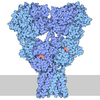
PDBj







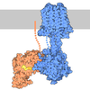



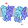











- Assembly
| Deposited unit | 
|
|---|---|
| 1 |
|
-Components
-Guanine nucleotide-binding protein ... , 3 types, 3 molecules CEG
| #2: Protein | Mass: 7861.143 Da / Num. of mol.: 1 Source method: isolated from a genetically manipulated source Source: (gene. exp.) Homo sapiens (human) / Gene: GNG2 / Production host: Homo sapiens (human) / References: UniProt: P59768 |
|---|---|
| #4: Protein | Mass: 40415.031 Da / Num. of mol.: 1 Source method: isolated from a genetically manipulated source Source: (gene. exp.) Homo sapiens (human) / Gene: GNAI1 / Production host: Homo sapiens (human)References: UniProt: P63096, Hydrolases; Acting on acid anhydrides; Acting on GTP to facilitate cellular and subcellular movement |
| #5: Protein | Mass: 39286.891 Da / Num. of mol.: 1 Source method: isolated from a genetically manipulated source Source: (gene. exp.) Homo sapiens (human) / Gene: GNB1 / Production host: Homo sapiens (human) / References: UniProt: P62873 |
-Protein / Antibody , 2 types, 3 molecules ABD
| #1: Protein | Mass: 94195.492 Da / Num. of mol.: 2 Source method: isolated from a genetically manipulated source Source: (gene. exp.) Homo sapiens (human) / Gene: GRM8, GPRC1H, MGLUR8 / Production host: Homo sapiens (human) / References: UniProt: O00222#3: Antibody | | Mass: 27784.896 Da / Num. of mol.: 1 Source method: isolated from a genetically manipulated source Source: (gene. exp.) Mus musculus (house mouse) / Production host: Homo sapiens (human) |
|---|
-Non-polymers , 2 types, 3 molecules 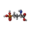
| #6: Chemical | 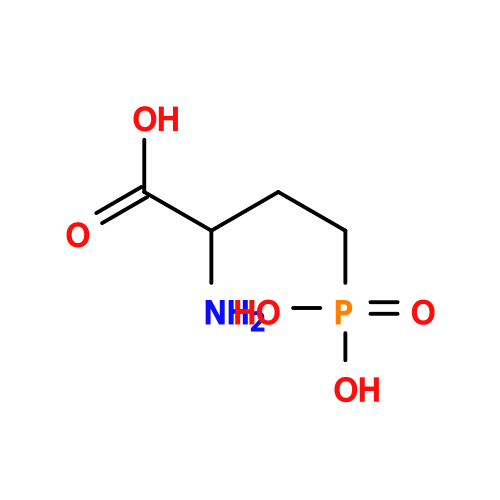 Mass: 183.100 Da / Num. of mol.: 2 / Source method: obtained synthetically / Formula: C4H10NO5P / Feature type: SUBJECT OF INVESTIGATION / Comment: agonist*YM Mass: 183.100 Da / Num. of mol.: 2 / Source method: obtained synthetically / Formula: C4H10NO5P / Feature type: SUBJECT OF INVESTIGATION / Comment: agonist*YM#7: Chemical | ChemComp-A1BWG / ( | Mass: 357.278 Da / Num. of mol.: 1 / Source method: obtained synthetically / Formula: C16H12F5N3O / Feature type: SUBJECT OF INVESTIGATION |
|---|
-Details
| Has ligand of interest | Y |
|---|---|
| Has protein modification | Y |
-Experimental details
-Experiment
| Experiment | Method: ELECTRON MICROSCOPY |
|---|---|
| EM experiment | Aggregation state: PARTICLE / 3D reconstruction method: single particle reconstruction |
- Sample preparation
| Component |
| ||||||||||||||||||||||||
|---|---|---|---|---|---|---|---|---|---|---|---|---|---|---|---|---|---|---|---|---|---|---|---|---|---|
| Molecular weight |
| ||||||||||||||||||||||||
| Source (natural) |
| ||||||||||||||||||||||||
| Source (recombinant) |
| ||||||||||||||||||||||||
| Buffer solution | pH: 7.5 | ||||||||||||||||||||||||
| Specimen | Conc.: 4.5 mg/ml / Embedding applied: NO / Shadowing applied: NO / Staining applied: NO / Vitrification applied: YES | ||||||||||||||||||||||||
| Vitrification | Instrument: FEI VITROBOT MARK IV / Cryogen name: ETHANE |
- Electron microscopy imaging
| Microscopy | Model: TFS GLACIOS |
|---|---|
| Electron gun | Electron source:  FIELD EMISSION GUN / Accelerating voltage: 200 kV / Illumination mode: FLOOD BEAM FIELD EMISSION GUN / Accelerating voltage: 200 kV / Illumination mode: FLOOD BEAM |
| Electron lens | Mode: BRIGHT FIELD / Nominal defocus max: 2000 nm / Nominal defocus min: 700 nm |
| Image recording | Electron dose: 48 e/Å2 / Film or detector model: FEI FALCON IV (4k x 4k) |
- Processing
| EM software |
| |||||||||
|---|---|---|---|---|---|---|---|---|---|---|
| CTF correction | Type: PHASE FLIPPING AND AMPLITUDE CORRECTION | |||||||||
| 3D reconstruction | Resolution: 6.18 Å / Resolution method: FSC 0.143 CUT-OFF / Num. of particles: 142839 / Symmetry type: POINT |
