 Movie
Movie Controller


+ Open data

- Basic information
| Entry | Database: PDB / ID: 9xzs | ||||||||||||||||||||||||||||||
|---|---|---|---|---|---|---|---|---|---|---|---|---|---|---|---|---|---|---|---|---|---|---|---|---|---|---|---|---|---|---|---|
| Title | Trm10-tRNA complex (Two Trm10 monomers bound to one tRNA) | ||||||||||||||||||||||||||||||
 Components Components |
| ||||||||||||||||||||||||||||||
 Keywords Keywords | RNA BINDING PROTEIN/RNA / SPOUT methyltransferase / tRNA / methylation / RNA BINDING PROTEIN / RNA BINDING PROTEIN-RNA complex | ||||||||||||||||||||||||||||||
| Function / homology |  Function and homology information Function and homology informationtRNA (guanine) methyltransferase activity / tRNA (guanine9-N1)-methyltransferase / tRNA (guanosine(9)-N1)-methyltransferase activity / tRNA N1-guanine methylation / tRNA modification / tRNA methylation / tRNA binding / nucleoplasm / nucleus / cytoplasm Similarity search - Function | ||||||||||||||||||||||||||||||
| Biological species |  | ||||||||||||||||||||||||||||||
| Method | ELECTRON MICROSCOPY / single particle reconstruction / cryo EM / Resolution: 3.89 Å | ||||||||||||||||||||||||||||||
 Authors Authors | Nandi, S. / Strassler, S.E. / Conn, G.L. | ||||||||||||||||||||||||||||||
| Funding support |  United States, 1items United States, 1items
| ||||||||||||||||||||||||||||||
 Citation Citation | Journal: bioRxiv / Year: 2025 Title: Molecular basis of tRNA substrate recognition and modification by the atypical SPOUT methyltransferase Trm10. Authors: Suparno Nandi / Sarah E Strassler / Debayan Dey / Aiswarya Krishnamohan / George M Harris / Lindsay R Comstock / Jane E Jackman / Graeme L Conn / Abstract: The evolutionarily conserved methyltransferase Trm10 modifies the N1 position of guanosine 9 (G9) in some tRNAs, but how the enzyme recognizes and modifies its substrate tRNAs remains unclear. Here, ...The evolutionarily conserved methyltransferase Trm10 modifies the N1 position of guanosine 9 (G9) in some tRNAs, but how the enzyme recognizes and modifies its substrate tRNAs remains unclear. Here, we used an S-adenosyl-L-methionine (SAM) analog to trap the Trm10-tRNA complex and enable determination of its structure in a post-catalytic state by cryogenic electron microscopy (cryo-EM). We observed three distinct complexes: two with a single Trm10 bound to tRNA that differ in their tRNA acceptor stem orientation ("closed" and "open") and a minor population with two Trm10s engaging the same tRNA. The monomeric complexes reveal a positively charged surface that guides the G9 into the catalytic site with key conserved residues forming "pincer"-like interactions that stabilize the flipped methylated nucleotide. In the open tRNA conformation, the acceptor stem is rotated away from the enzyme, weakening the tRNA-protein contacts, consistent with a product-release conformation. The dimeric complex, which is supported by tRNA-dependent protein crosslinking, reveals one Trm10 positioned similarly to the monomeric complexes and engaged with G9, while the other Trm10 contacts distal tRNA regions, suggesting a potential role in facilitating a key conformational transition during the process of catalysis or modified tRNA release. Finally, molecular dynamics simulations comparing G9- and A9-containing complexes reveal that G9 is efficiently stabilized in the binding pocket unlike A9, identifying the structural basis for guanosine selectivity. Overall, these findings reveal the structural determinants of G9-specific tRNA methylation by Trm10 and suggest a unique mechanism of action among RNA-modifying SPOUT methyltransferases. | ||||||||||||||||||||||||||||||
| History |
|
- Structure visualization
| Structure viewer | Molecule: MolmilJmol/JSmol |
|---|
- Downloads & links
-Download
| PDBx/mmCIF format | 9xzs.cif.gz | 119.3 KB | Display | PDBx/mmCIF format |
|---|---|---|---|---|
| PDB format | pdb9xzs.ent.gz | Display | PDB format | |
| PDBx/mmJSON format | 9xzs.json.gz | Tree view | PDBx/mmJSON format | |
| Others |  Other downloads Other downloads |
-Validation report
| Arichive directory | https://data.pdbj.org/pub/pdb/validation_reports/xz/9xzsftp://data.pdbj.org/pub/pdb/validation_reports/xz/9xzs | HTTPS FTP |
|---|
-Related structure data
| Related structure data |  72370MC  9xzqC  9xzrC M: map data used to model this data C: citing same article ( |
|---|---|
| Similar structure data |


-Links
 PDBj
PDBj


- Assembly
| Deposited unit | 
|
|---|---|
| 1 |
|
-Components
| #1: RNA chain | Mass: 22832.520 Da / Num. of mol.: 1 / Source method: obtained synthetically / Source: (synth.) | ||||||
|---|---|---|---|---|---|---|---|
| #2: Protein | Mass: 34583.582 Da / Num. of mol.: 2 Source method: isolated from a genetically manipulated source Source: (gene. exp.) Gene: TRM10, YOL093W, O0926 / Production host:  References: UniProt: Q12400, tRNA (guanine9-N1)-methyltransferase #3: Chemical | ChemComp-AN6 / | 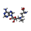  Type: L-peptide linking / Mass: 395.414 Da / Num. of mol.: 1 / Source method: obtained synthetically / Formula: C16H25N7O5 / Source: (synth.) Type: L-peptide linking / Mass: 395.414 Da / Num. of mol.: 1 / Source method: obtained synthetically / Formula: C16H25N7O5 / Source: (synth.) Has ligand of interest | N | Has protein modification | N | |
-Experimental details
-Experiment
| Experiment | Method: ELECTRON MICROSCOPY |
|---|---|
| EM experiment | Aggregation state: PARTICLE / 3D reconstruction method: single particle reconstruction |
- Sample preparation
| Component | Name: Ternary complex of two Trm10 molecules bound to tRNA / Type: COMPLEX / Entity ID: #1-#2 / Source: RECOMBINANT |
|---|---|
| Molecular weight | Experimental value: NO |
| Source (natural) | Organism: |
| Source (recombinant) | Organism: |
| Buffer solution | pH: 7.5 |
| Specimen | Embedding applied: NO / Shadowing applied: NO / Staining applied: NO / Vitrification applied: YES |
| Specimen support | Grid material: GOLD / Grid mesh size: 300 divisions/in. / Grid type: UltrAuFoil |
| Vitrification | Instrument: FEI VITROBOT MARK IV / Cryogen name: ETHANE / Humidity: 100 % / Chamber temperature: 298 K |
- Electron microscopy imaging
| Experimental equipment |  Model: Titan Krios / Image courtesy: FEI Company |
|---|---|
| Microscopy | Model: TFS KRIOS |
| Electron gun | Electron source:  FIELD EMISSION GUN / Accelerating voltage: 300 kV / Illumination mode: FLOOD BEAM FIELD EMISSION GUN / Accelerating voltage: 300 kV / Illumination mode: FLOOD BEAM |
| Electron lens | Mode: BRIGHT FIELD / Nominal defocus max: 2000 nm / Nominal defocus min: 800 nm / Cs: 2.7 mm |
| Image recording | Average exposure time: 2 sec. / Electron dose: 59.82 e/Å2 / Film or detector model: GATAN K3 BIOQUANTUM (6k x 4k) / Num. of real images: 26645 |
| Image scans | Width: 11520 / Height: 8184 |
- Processing
| EM software |
| ||||||||||||||||||||||||||||||||||||||||
|---|---|---|---|---|---|---|---|---|---|---|---|---|---|---|---|---|---|---|---|---|---|---|---|---|---|---|---|---|---|---|---|---|---|---|---|---|---|---|---|---|---|
| CTF correction | Type: PHASE FLIPPING AND AMPLITUDE CORRECTION | ||||||||||||||||||||||||||||||||||||||||
| Particle selection | Num. of particles selected: 14103903 | ||||||||||||||||||||||||||||||||||||||||
| 3D reconstruction | Resolution: 3.89 Å / Resolution method: FSC 0.143 CUT-OFF / Num. of particles: 156897 / Symmetry type: POINT | ||||||||||||||||||||||||||||||||||||||||
| Atomic model building | Protocol: FLEXIBLE FIT / Space: REAL | ||||||||||||||||||||||||||||||||||||||||
| Atomic model building | Details: chain B of the model is from PDB 4JWJ. Chain A was prepared using AlphaFold. Source name: Other / Type: integrative model |
