Movie
 Controller


[English] 日本語
 Yorodumi
- PDB-9svi: Cryo-EM structure of CRBN bound to 1-[1-(4-piperidyl)indol-4-yl]h... -
+ Open data

- Basic information
| Entry | Database: PDB / ID: 9svi | |||||||||
|---|---|---|---|---|---|---|---|---|---|---|
| Title | Cryo-EM structure of CRBN bound to 1-[1-(4-piperidyl)indol-4-yl]hexahydropyrimidine-2,4-dione in the open conformation | |||||||||
 Components |
| |||||||||
 Keywords | LIGASE / E3 ubiquitin ligase | |||||||||
| Function / homology |  Function and homology information negative regulation of monoatomic ion transmembrane transport / positive regulation by virus of viral protein levels in host cell / spindle assembly involved in female meiosis / epigenetic programming in the zygotic pronuclei / UV-damage excision repair / biological process involved in interaction with symbiont / regulation of mitotic cytokinesis / regulation of mitotic cell cycle phase transition / regulation of miRNA-mediated gene silencing / regulation of natural killer cell activation ...negative regulation of monoatomic ion transmembrane transport / positive regulation by virus of viral protein levels in host cell / spindle assembly involved in female meiosis / epigenetic programming in the zygotic pronuclei / UV-damage excision repair / biological process involved in interaction with symbiont / regulation of mitotic cytokinesis / regulation of mitotic cell cycle phase transition / regulation of miRNA-mediated gene silencing / regulation of natural killer cell activation / WD40-repeat domain binding / nucleotide-excision repair complex / regulation of cell cycle phase transition / Cul4-RING E3 ubiquitin ligase complex / locomotory exploration behavior / regulation of stem cell population maintenance / ubiquitin ligase complex scaffold activity / negative regulation of adipose tissue development / Cul4B-RING E3 ubiquitin ligase complex / regulation of cellular response to stress / limb development / viral release from host cell / Cul4A-RING E3 ubiquitin ligase complex / cullin family protein binding / positive regulation of Wnt signaling pathway / negative regulation of protein-containing complex assembly / regulation of DNA-templated DNA replication initiation / positive regulation of viral genome replication / positive regulation of gluconeogenesis / regulation of embryonic development / replication fork processing / proteasomal protein catabolic process / epigenetic regulation of gene expression / positive regulation of protein-containing complex assembly / nucleotide-excision repair / regulation of autophagy / Recognition of DNA damage by PCNA-containing replication complex / regulation of circadian rhythm / cell population proliferation / DNA Damage Recognition in GG-NER / Transcription-Coupled Nucleotide Excision Repair (TC-NER) / Formation of TC-NER Pre-Incision Complex / Dual Incision in GG-NER / Formation of Incision Complex in GG-NER / cellular response to UV / Dual incision in TC-NER / positive regulation of protein catabolic process / Gap-filling DNA repair synthesis and ligation in TC-NER / site of double-strand break / regulation of cell population proliferation / Neddylation / spermatogenesis / ubiquitin-dependent protein catabolic process / Potential therapeutics for SARS / proteasome-mediated ubiquitin-dependent protein catabolic process / damaged DNA binding / regulation of apoptotic process / transmembrane transporter binding / chromosome, telomeric region / protein-macromolecule adaptor activity / protein ubiquitination / DNA repair / DNA damage response / nucleolus / protein-containing complex binding / perinuclear region of cytoplasm / protein-containing complex / DNA binding / extracellular exosome / nucleoplasm / extracellular region / membrane / nucleus / cytosol / cytoplasm Similarity search - Function | |||||||||
| Biological species |  Homo sapiens (human) | |||||||||
| Method | ELECTRON MICROSCOPY / single particle reconstruction / cryo EM / Resolution: 3.33 Å | |||||||||
 Authors | Cowan, A.D. / Rutter, Z.J. / McAulay, K. / Ciulli, A. | |||||||||
| Funding support |  Germany, 1items
| |||||||||
 Citation | Journal: To Be Published Title: Structural and Biophysical Methods for the Assessment of Cereblon Binders for PROTACs Authors: Rutter, Z.J. / Cowan, A.D. / McAulay, K. / Ciulli, A. | |||||||||
| History |
|
- Structure visualization
| Structure viewer | Molecule: MolmilJmol/JSmol |
|---|
- Downloads & links
-Download
| PDBx/mmCIF format | 9svi.cif.gz | 452.8 KB | Display | PDBx/mmCIF format |
|---|---|---|---|---|
| PDB format | pdb9svi.ent.gz | Display | PDB format | |
| PDBx/mmJSON format | 9svi.json.gz | Tree view | PDBx/mmJSON format | |
| Others |  Other downloads |
-Validation report
| Arichive directory | https://data.pdbj.org/pub/pdb/validation_reports/sv/9sviftp://data.pdbj.org/pub/pdb/validation_reports/sv/9svi | HTTPS FTP |
|---|
-Related structure data
| Related structure data |  55259MC  9sunC  9svgC  9svhC C: citing same article ( M: map data used to model this data |
|---|---|
| Similar structure data |


-Links
 PDBj
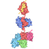
- Assembly
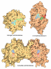
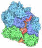
| Deposited unit | 
|
|---|---|
| 1 |
|
-Components
| #1: Protein | Mass: 93347.078 Da / Num. of mol.: 1 Source method: isolated from a genetically manipulated source Source: (gene. exp.) Homo sapiens (human) / Gene: DDB1, XAP1 / Production host:  Trichoplusia ni (cabbage looper) / References: UniProt: Q16531 |
|---|---|
| #2: Protein | Mass: 50559.559 Da / Num. of mol.: 1 Source method: isolated from a genetically manipulated source Source: (gene. exp.) Homo sapiens (human) / Gene: CRBN, AD-006 / Production host: Trichoplusia ni (cabbage looper) / References: UniProt: Q96SW2 |
| #3: Chemical | ChemComp-ZN /   Mass: 65.409 Da / Num. of mol.: 1 / Source method: obtained synthetically / Formula: Zn |
| #4: Chemical | ChemComp-A1JP8 / Mass: 312.366 Da / Num. of mol.: 1 / Source method: obtained synthetically / Formula: C17H20N4O2 / Feature type: SUBJECT OF INVESTIGATION |
| Has ligand of interest | Y |
| Has protein modification | N |
-Experimental details
-Experiment
| Experiment | Method: ELECTRON MICROSCOPY |
|---|---|
| EM experiment | Aggregation state: PARTICLE / 3D reconstruction method: single particle reconstruction |
- Sample preparation
| Component | Name: Complex of CRL4 E3 adapter-receptor pair DDB1 and CRBN bound to dihydrouracil ligand Type: COMPLEX / Entity ID: #1-#2 / Source: RECOMBINANT | |||||||||||||||||||||||||
|---|---|---|---|---|---|---|---|---|---|---|---|---|---|---|---|---|---|---|---|---|---|---|---|---|---|---|
| Molecular weight | Value: 0.14377612 MDa / Experimental value: NO | |||||||||||||||||||||||||
| Source (natural) | Organism: Homo sapiens (human) | |||||||||||||||||||||||||
| Source (recombinant) | Organism: Trichoplusia ni (cabbage looper) | |||||||||||||||||||||||||
| Buffer solution | pH: 7.4 | |||||||||||||||||||||||||
| Buffer component |
| |||||||||||||||||||||||||
| Specimen | Conc.: 0.72 mg/ml / Embedding applied: NO / Shadowing applied: NO / Staining applied: NO / Vitrification applied: YES | |||||||||||||||||||||||||
| Specimen support | Details: 35 mA / Grid material: GOLD / Grid mesh size: 300 divisions/in. / Grid type: UltrAuFoil R1.2/1.3 | |||||||||||||||||||||||||
| Vitrification | Cryogen name: ETHANE / Humidity: 100 % / Chamber temperature: 277 K |
- Electron microscopy imaging
| Microscopy | Model: TFS GLACIOS |
|---|---|
| Electron gun | Electron source:  FIELD EMISSION GUN / Accelerating voltage: 200 kV / Illumination mode: FLOOD BEAM |
| Electron lens | Mode: BRIGHT FIELD / Nominal magnification: 190000 X / Nominal defocus max: 3200 nm / Nominal defocus min: 1700 nm |
| Specimen holder | Cryogen: NITROGEN / Specimen holder model: FEI TITAN KRIOS AUTOGRID HOLDER |
| Image recording | Average exposure time: 2.35 sec. / Electron dose: 21.94 e/Å2 / Film or detector model: TFS FALCON 4i (4k x 4k) / Num. of grids imaged: 1 / Num. of real images: 3752 |
- Processing
| EM software |
| ||||||||||||||||||||||||||||||||||||
|---|---|---|---|---|---|---|---|---|---|---|---|---|---|---|---|---|---|---|---|---|---|---|---|---|---|---|---|---|---|---|---|---|---|---|---|---|---|
| CTF correction | Details: Patch CTF / Type: PHASE FLIPPING AND AMPLITUDE CORRECTION | ||||||||||||||||||||||||||||||||||||
| 3D reconstruction | Resolution: 3.33 Å / Resolution method: FSC 0.143 CUT-OFF / Num. of particles: 86485 Details: cryoSPARC non-uniform refinement was used for the final reconstruction of the consensus map, and cryoSPARC local refinement was used for the locally refined map of the thalidomide binding domain Symmetry type: POINT | ||||||||||||||||||||||||||||||||||||
| Atomic model building | Protocol: FLEXIBLE FIT / Space: REAL Details: Initial fitting was done in ChimeraX, followed by flexible fitting in Coot. The model was refined with iterative rounds of building in Coot and refinement in PHENIX real space refine | ||||||||||||||||||||||||||||||||||||
| Atomic model building |
| ||||||||||||||||||||||||||||||||||||
| Refinement | Cross valid method: NONE Stereochemistry target values: GeoStd + Monomer Library + CDL v1.2 | ||||||||||||||||||||||||||||||||||||
| Displacement parameters | Biso mean: 51.39 Å2 | ||||||||||||||||||||||||||||||||||||
| Refine LS restraints |
|