Movie
Movie Controller

[English] 日本語
 Yorodumi
- EMDB-54644: Locally refined map of CRBN TBD bound to spirocyclic ligand in th... -
+ Open data

- Basic information
| Entry |  | |||||||||
|---|---|---|---|---|---|---|---|---|---|---|
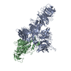
| Title | Locally refined map of CRBN TBD bound to spirocyclic ligand in the open conformation | |||||||||
 Map data Map data | Sharpened focused map | |||||||||
 Sample Sample |
| |||||||||
 Keywords Keywords | E3 ubiquitin ligase / LIGASE | |||||||||
| Function / homology |  Function and homology information Function and homology informationnegative regulation of monoatomic ion transmembrane transport / locomotory exploration behavior / Cul4A-RING E3 ubiquitin ligase complex / limb development / positive regulation of Wnt signaling pathway / negative regulation of protein-containing complex assembly / positive regulation of protein-containing complex assembly / Potential therapeutics for SARS / proteasome-mediated ubiquitin-dependent protein catabolic process / transmembrane transporter binding ...negative regulation of monoatomic ion transmembrane transport / locomotory exploration behavior / Cul4A-RING E3 ubiquitin ligase complex / limb development / positive regulation of Wnt signaling pathway / negative regulation of protein-containing complex assembly / positive regulation of protein-containing complex assembly / Potential therapeutics for SARS / proteasome-mediated ubiquitin-dependent protein catabolic process / transmembrane transporter binding / protein ubiquitination / perinuclear region of cytoplasm / membrane / metal ion binding / nucleus / cytosol / cytoplasm Similarity search - Function | |||||||||
| Biological species |  Homo sapiens (human) Homo sapiens (human) | |||||||||
| Method | single particle reconstruction / cryo EM / Resolution: 3.46 Å | |||||||||
 Authors Authors | Cowan AD / Rutter ZJ / McAulay K / Ciulli A | |||||||||
| Funding support |  Germany, 1 items Germany, 1 items
| |||||||||
 Citation Citation | Journal: Protein Sci / Year: 2023 Title: UCSF ChimeraX: Tools for structure building and analysis. Authors: Elaine C Meng / Thomas D Goddard / Eric F Pettersen / Greg S Couch / Zach J Pearson / John H Morris / Thomas E Ferrin /  Abstract: Advances in computational tools for atomic model building are leading to accurate models of large molecular assemblies seen in electron microscopy, often at challenging resolutions of 3-4 Å. We ...Advances in computational tools for atomic model building are leading to accurate models of large molecular assemblies seen in electron microscopy, often at challenging resolutions of 3-4 Å. We describe new methods in the UCSF ChimeraX molecular modeling package that take advantage of machine-learning structure predictions, provide likelihood-based fitting in maps, and compute per-residue scores to identify modeling errors. Additional model-building tools assist analysis of mutations, post-translational modifications, and interactions with ligands. We present the latest ChimeraX model-building capabilities, including several community-developed extensions. ChimeraX is available free of charge for noncommercial use at https://www.rbvi.ucsf.edu/chimerax. | |||||||||
| History |
|
- Structure visualization
| Supplemental images |
|---|

- Downloads & links
-EMDB archive
| Map data | emd_54644.map.gz | 167.7 MB | EMDB map data format | |
|---|---|---|---|---|
| Header (meta data) | emd-54644-v30.xmlemd-54644.xml | 23.7 KB 23.7 KB | Display Display | EMDB header |
| FSC (resolution estimation) | emd_54644_fsc.xml | 12 KB | Display | FSC data file |
| Images |  emd_54644.png emd_54644.png | 46.8 KB | ||
| Masks | emd_54644_msk_1.map | 178 MB | Mask map | |
| Filedesc metadata | emd-54644.cif.gz | 6.2 KB | ||
| Others | emd_54644_additional_1.map.gzemd_54644_half_map_1.map.gzemd_54644_half_map_2.map.gz | 88.2 MB 165.3 MB 165.3 MB | ||
| Archive directory |  http://ftp.pdbj.org/pub/emdb/structures/EMD-54644ftp://ftp.pdbj.org/pub/emdb/structures/EMD-54644 http://ftp.pdbj.org/pub/emdb/structures/EMD-54644ftp://ftp.pdbj.org/pub/emdb/structures/EMD-54644 | HTTPS FTP |
-Related structure data
| Related structure data | C: citing same article ( |
|---|---|
| Similar structure data |

-Links
| EMDB pages | EMDB (EBI/PDBe) / EMDataResource |
|---|---|
| Related items in Molecule of the Month |
-Map
| File | Download / File: emd_54644.map.gz / Format: CCP4 / Size: 178 MB / Type: IMAGE STORED AS FLOATING POINT NUMBER (4 BYTES) | ||||||||||||||||||||||||||||||||||||
|---|---|---|---|---|---|---|---|---|---|---|---|---|---|---|---|---|---|---|---|---|---|---|---|---|---|---|---|---|---|---|---|---|---|---|---|---|---|
| Annotation | Sharpened focused map | ||||||||||||||||||||||||||||||||||||
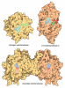
| Projections & slices | Image control
Images are generated by Spider. | ||||||||||||||||||||||||||||||||||||
| Voxel size | X=Y=Z: 0.749 Å | ||||||||||||||||||||||||||||||||||||
| Density |
| ||||||||||||||||||||||||||||||||||||
| Symmetry | Space group: 1 | ||||||||||||||||||||||||||||||||||||
| Details | EMDB XML:
|
 Z (Sec.)
 Y (Row.)
 X (Col.)


-Supplemental data
-Mask #1
| File | emd_54644_msk_1.map | ||||||||||||
|---|---|---|---|---|---|---|---|---|---|---|---|---|---|
| Projections & Slices |
| ||||||||||||
| Density Histograms |


-Additional map: Unsharpened focused map
| File | emd_54644_additional_1.map | ||||||||||||
|---|---|---|---|---|---|---|---|---|---|---|---|---|---|
| Annotation | Unsharpened focused map | ||||||||||||
| Projections & Slices |
| ||||||||||||
| Density Histograms |


-Half map: Focused half map A
| File | emd_54644_half_map_1.map | ||||||||||||
|---|---|---|---|---|---|---|---|---|---|---|---|---|---|
| Annotation | Focused half map A | ||||||||||||
| Projections & Slices |
| ||||||||||||
| Density Histograms |


-Half map: #1
| File | emd_54644_half_map_2.map | ||||||||||||
|---|---|---|---|---|---|---|---|---|---|---|---|---|---|
| Projections & Slices |
| ||||||||||||
| Density Histograms |


- Sample components
-Entire : Complex of CRL4 E3 adapter-receptor pair DDB1 and CRBN bound to s...
| Entire | Name: Complex of CRL4 E3 adapter-receptor pair DDB1 and CRBN bound to spirocyclic ligand |
|---|---|
| Components |
|
-Supramolecule #1: Complex of CRL4 E3 adapter-receptor pair DDB1 and CRBN bound to s...
| Supramolecule | Name: Complex of CRL4 E3 adapter-receptor pair DDB1 and CRBN bound to spirocyclic ligand type: complex / ID: 1 / Parent: 0 / Macromolecule list: all Details: Locally refined map of CRBN thalidomide binding domain bound to spirocyclic ligand |
|---|---|
| Source (natural) | Organism: Homo sapiens (human) |
| Molecular weight | Theoretical: 143.77612 KDa |
-Macromolecule #1: Cereblon
| Macromolecule | Name: Cereblon / type: protein_or_peptide / ID: 1 / Enantiomer: LEVO |
|---|---|
| Source (natural) | Organism: Homo sapiens (human) |
| Recombinant expression | Organism:  Trichoplusia ni (cabbage looper) Trichoplusia ni (cabbage looper) |
| Sequence | String: MAGEGDQQDA AHNMGNHLPL LPAESEEEDE MEVEDQDSKE AKKPNIINFD TSLPTSHTYL GADMEEFHGR TLHDDDSCQV IPVLPQVMM ILIPGQTLPL QLFHPQEVSM VRNLIQKDRT FAVLAYSNVQ EREAQFGTTA EIYAYREEQD FGIEIVKVKA I GRQRFKVL ...String: MAGEGDQQDA AHNMGNHLPL LPAESEEEDE MEVEDQDSKE AKKPNIINFD TSLPTSHTYL GADMEEFHGR TLHDDDSCQV IPVLPQVMM ILIPGQTLPL QLFHPQEVSM VRNLIQKDRT FAVLAYSNVQ EREAQFGTTA EIYAYREEQD FGIEIVKVKA I GRQRFKVL ELRTQSDGIQ QAKVQILPEC VLPSTMSAVQ LESLNKCQIF PSKPVSREDQ CSYKWWQKYQ KRKFHCANLT SW PRWLYSL YDAETLMDRI KKQLREWDEN LKDDSLPSNP IDFSYRVAAC LPIDDVLRIQ LLKIGSAIQR LRCELDIMNK CTS LCCKQC QETEITTKNE IFSLSLCGPM AAYVNPHGYV GETLTVYKAC NLNLIGRPST EHSWFPGYAW TVAQCKICAS HIGW KFTAT KKDMSPQKFW GLTRSALLPT IPDTEDEISP DKVILCL UniProtKB: Protein cereblon |
-Experimental details
-Structure determination
| Method | cryo EM |
|---|---|
 Processing Processing | single particle reconstruction |
| Aggregation state | particle |
-Sample preparation
| Concentration | 7.2 mg/mL | |||||||||||||||
|---|---|---|---|---|---|---|---|---|---|---|---|---|---|---|---|---|
| Buffer | pH: 7.5 Component:
| |||||||||||||||
| Vitrification | Cryogen name: ETHANE / Chamber humidity: 100 % / Chamber temperature: 277 K / Instrument: FEI VITROBOT MARK IV |
- Electron microscopy
| Microscope | TFS KRIOS |
|---|---|
| Image recording | Film or detector model: TFS FALCON 4i (4k x 4k) / Average electron dose: 50.0 e/Å2 |
| Electron beam | Acceleration voltage: 300 kV / Electron source:  FIELD EMISSION GUN FIELD EMISSION GUN |
| Electron optics | Illumination mode: FLOOD BEAM / Imaging mode: BRIGHT FIELD / Nominal defocus max: 1.8 µm / Nominal defocus min: 0.8 µm |
| Sample stage | Specimen holder model: FEI TITAN KRIOS AUTOGRID HOLDER |
| Experimental equipment |  Model: Titan Krios / Image courtesy: FEI Company |
+Image processing

-Atomic model buiding 1
| Initial model | PDB ID: Chain - Source name: PDB / Chain - Initial model type: experimental model |
|---|---|
| Details | Initial fitting was done in ChimeraX, followed by flexible fitting in Coot. The model was refined with iterative rounds of building in Coot and refinement in PHENIX real space refine |
| Refinement | Space: REAL / Protocol: FLEXIBLE FIT |