 Movie
Movie Controller


+ Open data

- Basic information
| Entry | Database: PDB / ID: 9sk3 | |||||||||||||||||||||||||||||||||||||||
|---|---|---|---|---|---|---|---|---|---|---|---|---|---|---|---|---|---|---|---|---|---|---|---|---|---|---|---|---|---|---|---|---|---|---|---|---|---|---|---|---|
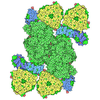
| Title | Trimeric photosystem I of Acaryochloris marina NIES-2412 | |||||||||||||||||||||||||||||||||||||||
 Components Components | (Photosystem I protein ...) x 12 | |||||||||||||||||||||||||||||||||||||||
 Keywords Keywords | ELECTRON TRANSPORT / Chlorophyll d / photosystem I | |||||||||||||||||||||||||||||||||||||||
| Function / homology | : / CHLOROPHYLL D / CHLOROPHYLL D ISOMER / 1,2-DIPALMITOYL-PHOSPHATIDYL-GLYCEROLE / 1,2-DISTEAROYL-MONOGALACTOSYL-DIGLYCERIDE / PHEOPHYTIN A / PHYLLOQUINONE / IRON/SULFUR CLUSTER Function and homology information Function and homology information | |||||||||||||||||||||||||||||||||||||||
| Biological species |  Acaryochloris marina NIES-2412 (bacteria) Acaryochloris marina NIES-2412 (bacteria) | |||||||||||||||||||||||||||||||||||||||
| Method | ELECTRON MICROSCOPY / single particle reconstruction / cryo EM / Resolution: 2.63 Å | |||||||||||||||||||||||||||||||||||||||
 Authors Authors | Leong, H.F. / Consoli, G. | |||||||||||||||||||||||||||||||||||||||
| Funding support |  Netherlands, European Union, Netherlands, European Union,  United Kingdom, 12items United Kingdom, 12items
| |||||||||||||||||||||||||||||||||||||||
 Citation Citation | Journal: Sci Adv / Year: 2026 Title: Far-red chlorophyll d clusters extend photosystem I absorption toward the red limit. Authors: Thomas J Oliver / Eduard Elias / Giovanni Consoli / Ho Fong Leong / Violeta Cordón-Preciado / Andrea Fantuzzi / Tanai Cardona / A William Rutherford / Roberta Croce / Abstract: Oxygenic photosynthesis is usually limited to visible light, but the marine cyanobacterium pushes this boundary by harvesting far-red photons with chlorophyll d. The best-studied strain, MBIC11017, ...Oxygenic photosynthesis is usually limited to visible light, but the marine cyanobacterium pushes this boundary by harvesting far-red photons with chlorophyll d. The best-studied strain, MBIC11017, unexpectedly lacks low-energy chlorophylls ("red forms") in photosystem I, limiting absorption beyond 740 nanometers. Here, we show that another strain, NIES-2412, has evolved a strategy to absorb far-red photons up to 760 nanometers. Combining time-resolved fluorescence spectroscopy with cryo-electron microscopy at 2.64-angstrom resolution, we identify two distinct classes of chlorophyll d red forms in its photosystem I. One class originates from classical charge-transfer-exciton mixing, while the other arises purely from excitonic interactions. Mapping all 96 chlorophylls d reveals the precise pigments responsible for these far-red states. We also uncover a previously unreported subunit, PsaX2, which stabilizes the photosystem I complex and shapes pigment geometry and energetics to enable the formation of red forms. Last, we show that the protein modifications responsible for binding and tuning these red forms are widespread across the genus but not within the model MBIC11017 strain. Far-red photons lie close to the energetic limit of oxygenic photosynthesis; their efficient use therefore requires fine-tuning of the photosynthetic machinery. To our knowledge, our findings provide the structural and mechanistic basis of one of the most red-shifted photosystem I complexes identified to date, highlighting a distinct adaptive strategy in far-red light environments and offering design principles for extending photosynthesis in crops into the infrared. #1: Journal: Protein Sci / Year: 2018 Title: UCSF ChimeraX: Meeting modern challenges in visualization and analysis. Authors: Thomas D Goddard / Conrad C Huang / Elaine C Meng / Eric F Pettersen / Gregory S Couch / John H Morris / Thomas E Ferrin /  Abstract: UCSF ChimeraX is next-generation software for the visualization and analysis of molecular structures, density maps, 3D microscopy, and associated data. It addresses challenges in the size, scope, and ...UCSF ChimeraX is next-generation software for the visualization and analysis of molecular structures, density maps, 3D microscopy, and associated data. It addresses challenges in the size, scope, and disparate types of data attendant with cutting-edge experimental methods, while providing advanced options for high-quality rendering (interactive ambient occlusion, reliable molecular surface calculations, etc.) and professional approaches to software design and distribution. This article highlights some specific advances in the areas of visualization and usability, performance, and extensibility. ChimeraX is free for noncommercial use and is available from http://www.rbvi.ucsf.edu/chimerax/ for Windows, Mac, and Linux. | |||||||||||||||||||||||||||||||||||||||
| History |
|
- Structure visualization
| Structure viewer | Molecule: MolmilJmol/JSmol |
|---|
- Downloads & links
-Download
| PDBx/mmCIF format | 9sk3.cif.gz | 1.7 MB | Display | PDBx/mmCIF format |
|---|---|---|---|---|
| PDB format | pdb9sk3.ent.gz | Display | PDB format | |
| PDBx/mmJSON format | 9sk3.json.gz | Tree view | PDBx/mmJSON format | |
| Others |  Other downloads Other downloads |
-Validation report
| Arichive directory | https://data.pdbj.org/pub/pdb/validation_reports/sk/9sk3ftp://data.pdbj.org/pub/pdb/validation_reports/sk/9sk3 | HTTPS FTP |
|---|
-Related structure data
| Related structure data |  54956MC  9s6pC M: map data used to model this data C: citing same article ( |
|---|---|
| Similar structure data | |
| Other databases |
|

-Links
 PDBj
PDBj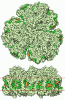

- Assembly
| Deposited unit | 
|
|---|---|
| 1 |
|
-Components
-Photosystem I protein ... , 12 types, 36 molecules aAbAcAaBbBcBaCbCcCaDbDcDaEbEcEaFbFcFaIbIcIaJbJcJaKbKcKaLbLcL...
| #1: Protein | Mass: 83573.766 Da / Num. of mol.: 3 / Source method: isolated from a natural source / Source: (natural) Acaryochloris marina NIES-2412 (bacteria)#2: Protein | Mass: 82083.172 Da / Num. of mol.: 3 / Source method: isolated from a natural source / Source: (natural) Acaryochloris marina NIES-2412 (bacteria)#3: Protein | Mass: 8825.207 Da / Num. of mol.: 3 / Source method: isolated from a natural source / Source: (natural) Acaryochloris marina NIES-2412 (bacteria)#4: Protein | Mass: 15125.150 Da / Num. of mol.: 3 / Source method: isolated from a natural source / Source: (natural) Acaryochloris marina NIES-2412 (bacteria)#5: Protein | Mass: 9350.654 Da / Num. of mol.: 3 / Source method: isolated from a natural source / Source: (natural) Acaryochloris marina NIES-2412 (bacteria)#6: Protein | Mass: 17691.422 Da / Num. of mol.: 3 / Source method: isolated from a natural source / Source: (natural) Acaryochloris marina NIES-2412 (bacteria)#7: Protein/peptide | Mass: 3648.337 Da / Num. of mol.: 3 / Source method: isolated from a natural source / Source: (natural) Acaryochloris marina NIES-2412 (bacteria)#8: Protein/peptide | Mass: 5457.358 Da / Num. of mol.: 3 / Source method: isolated from a natural source / Source: (natural) Acaryochloris marina NIES-2412 (bacteria)#9: Protein | Mass: 8894.473 Da / Num. of mol.: 3 / Source method: isolated from a natural source / Source: (natural) Acaryochloris marina NIES-2412 (bacteria)#10: Protein | Mass: 15588.004 Da / Num. of mol.: 3 / Source method: isolated from a natural source / Source: (natural) Acaryochloris marina NIES-2412 (bacteria)#11: Protein/peptide | Mass: 3204.925 Da / Num. of mol.: 3 / Source method: isolated from a natural source / Source: (natural) Acaryochloris marina NIES-2412 (bacteria)#12: Protein/peptide | Mass: 2973.575 Da / Num. of mol.: 3 / Source method: isolated from a natural source / Source: (natural) Acaryochloris marina NIES-2412 (bacteria) |
|---|
-Non-polymers , 9 types, 462 molecules 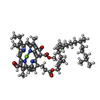

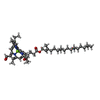
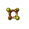

| #13: Chemical |  Mass: 895.462 Da / Num. of mol.: 3 / Source method: obtained synthetically / Formula: C54H70MgN4O6 Mass: 895.462 Da / Num. of mol.: 3 / Source method: obtained synthetically / Formula: C54H70MgN4O6#14: Chemical | ChemComp-PHO / 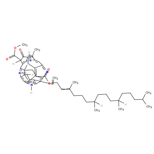 Mass: 871.200 Da / Num. of mol.: 6 / Source method: obtained synthetically / Formula: C55H74N4O5 Mass: 871.200 Da / Num. of mol.: 6 / Source method: obtained synthetically / Formula: C55H74N4O5#15: Chemical | ChemComp-CL7 /  Mass: 895.462 Da / Num. of mol.: 279 / Source method: obtained synthetically / Formula: C54H70MgN4O6 / Feature type: SUBJECT OF INVESTIGATION Mass: 895.462 Da / Num. of mol.: 279 / Source method: obtained synthetically / Formula: C54H70MgN4O6 / Feature type: SUBJECT OF INVESTIGATION#16: Chemical | ChemComp-PQN / 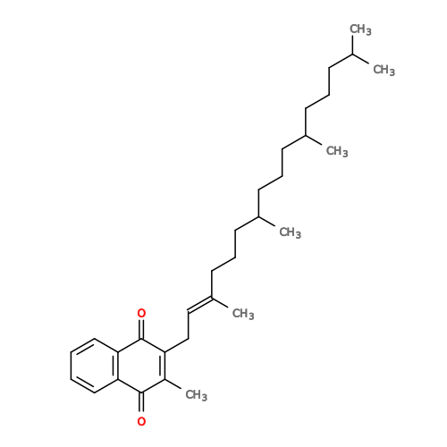 Mass: 450.696 Da / Num. of mol.: 6 / Source method: obtained synthetically / Formula: C31H46O2 Mass: 450.696 Da / Num. of mol.: 6 / Source method: obtained synthetically / Formula: C31H46O2#17: Chemical | ChemComp-A1JPJ / ( Mass: 536.873 Da / Num. of mol.: 57 / Source method: obtained synthetically / Formula: C40H56 #18: Chemical | ChemComp-LHG / 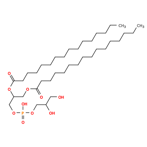 Mass: 722.970 Da / Num. of mol.: 6 / Source method: obtained synthetically / Formula: C38H75O10P / Comment: phospholipid*YM Mass: 722.970 Da / Num. of mol.: 6 / Source method: obtained synthetically / Formula: C38H75O10P / Comment: phospholipid*YM#19: Chemical | 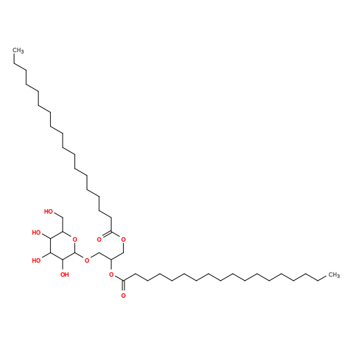 Mass: 787.158 Da / Num. of mol.: 3 / Source method: obtained synthetically / Formula: C45H86O10 Mass: 787.158 Da / Num. of mol.: 3 / Source method: obtained synthetically / Formula: C45H86O10#20: Chemical | ChemComp-SF4 / 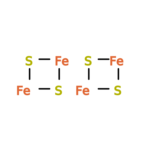 Mass: 351.640 Da / Num. of mol.: 9 / Source method: obtained synthetically / Formula: Fe4S4 Mass: 351.640 Da / Num. of mol.: 9 / Source method: obtained synthetically / Formula: Fe4S4#21: Water | ChemComp-HOH / | Mass: 18.015 Da / Num. of mol.: 93 / Source method: isolated from a natural source / Formula: H2O |
|---|
-Details
| Has ligand of interest | Y |
|---|---|
| Has protein modification | Y |
-Experimental details
-Experiment
| Experiment | Method: ELECTRON MICROSCOPY |
|---|---|
| EM experiment | Aggregation state: PARTICLE / 3D reconstruction method: single particle reconstruction |
- Sample preparation
| Component | Name: Trimeric PSI / Type: COMPLEX / Details: trimeric photosystem I from A. marina NIES - 2412 / Entity ID: #4-#5, #11-#12 / Source: NATURAL |
|---|---|
| Molecular weight | Value: 0.922 MDa / Experimental value: NO |
| Source (natural) | Organism: Acaryochloris marina (bacteria) / Strain: NIES-2412 |
| Buffer solution | pH: 6.5 Details: 50 mM MES, 1 M betaine monohydrate, 20 mM CaCl2, 5 mM MgCl2 and 10% (v/v) glycerol, pH 6.5 adjusted with NaOH |
| Specimen | Conc.: 1 mg/ml / Embedding applied: NO / Shadowing applied: NO / Staining applied: NO / Vitrification applied: YES |
| Specimen support | Grid material: GOLD / Grid mesh size: 300 divisions/in. / Grid type: UltrAuFoil |
| Vitrification | Instrument: FEI VITROBOT MARK IV / Cryogen name: ETHANE / Humidity: 95 % / Chamber temperature: 277 K |
- Electron microscopy imaging
| Experimental equipment |  Model: Titan Krios / Image courtesy: FEI Company |
|---|---|
| Microscopy | Model: TFS KRIOS |
| Electron gun | Electron source:  FIELD EMISSION GUN / Accelerating voltage: 300 kV / Illumination mode: SPOT SCAN FIELD EMISSION GUN / Accelerating voltage: 300 kV / Illumination mode: SPOT SCAN |
| Electron lens | Mode: BRIGHT FIELD / Nominal defocus max: 2000 nm / Nominal defocus min: 800 nm / Cs: 2.7 mm |
| Image recording | Electron dose: 40 e/Å2 / Film or detector model: GATAN K3 (6k x 4k) |
| Image scans | Width: 5120 / Height: 4096 |
- Processing
| EM software |
| ||||||||||||||||
|---|---|---|---|---|---|---|---|---|---|---|---|---|---|---|---|---|---|
| CTF correction | Type: PHASE FLIPPING AND AMPLITUDE CORRECTION | ||||||||||||||||
| 3D reconstruction | Resolution: 2.63 Å / Resolution method: FSC 0.143 CUT-OFF / Num. of particles: 151833 / Symmetry type: POINT |
