 Movie
Movie Controller

[English] 日本語
 Yorodumi
- PDB-9r22: Cryo-EM structure of the light-driven proton pump PsPR in deterge... -
+ Open data

- Basic information
| Entry | Database: PDB / ID: 9r22 | ||||||||||||
|---|---|---|---|---|---|---|---|---|---|---|---|---|---|
| Title | Cryo-EM structure of the light-driven proton pump PsPR in detergent micelle | ||||||||||||
 Components Components | microbial rhodopsin | ||||||||||||
 Keywords Keywords | MEMBRANE PROTEIN / proton transport / retinal / bioenergetics / proteorhodopsin / microbial rhodopsin | ||||||||||||
| Function / homology | EICOSANE / RETINAL Function and homology information Function and homology information | ||||||||||||
| Biological species |  Candidatus Pseudothioglobus sp. (bacteria) Candidatus Pseudothioglobus sp. (bacteria) | ||||||||||||
| Method | ELECTRON MICROSCOPY / single particle reconstruction / cryo EM / Resolution: 2.48 Å | ||||||||||||
 Authors Authors | Kovalev, K. / Stetsenko, A. / Guskov, A. | ||||||||||||
| Funding support |  Netherlands, Netherlands,  Germany, 3items Germany, 3items
| ||||||||||||
 Citation Citation | Journal: Structure / Year: 2025 Title: Structural basis for no retinal binding in flotillin-associated rhodopsins. Authors: Kirill Kovalev / Artem Stetsenko / Florian Trunk / Egor Marin / Jose M Haro-Moreno / Gerrit H U Lamm / Alexey Alekseev / Francisco Rodriguez-Valera / Thomas R Schneider / Josef Wachtveitl / Albert Guskov /  Abstract: Rhodopsins are light-sensitive membrane proteins capturing solar energy via a retinal cofactor covalently attached to a lysine residue. Several groups of rhodopsins lack the conserved lysine and ...Rhodopsins are light-sensitive membrane proteins capturing solar energy via a retinal cofactor covalently attached to a lysine residue. Several groups of rhodopsins lack the conserved lysine and showed no retinal binding. Recently, flotillin-associated rhodopsins (FArhodopsins or FARs) were identified and suggested to lack the retinal-binding pocket despite preserving the lysine residue in many members of the group. Here, we present cryoelectron microscopic (cryo-EM) structures of paralog FArhodopsin and proteorhodopsin from marine bacterium Pseudothioglobus, both forming pentamers similar to those of other microbial rhodopsins. We demonstrate no binding of retinal to the FArhodopsin despite preservation of the lysine residue and overall similarity of the protein fold and internal organization to those of the retinal-binding paralog. Mutational analysis confirmed that two amino acids, H84 and E120, prevent retinal binding within the FArhodopsin. Our work provides insights into the natural retinal loss in microbial rhodopsins and might contribute to the further understanding of FArhodopsins. | ||||||||||||
| History |
|
- Structure visualization
| Structure viewer | Molecule: MolmilJmol/JSmol |
|---|
- Downloads & links
-Download
| PDBx/mmCIF format | 9r22.cif.gz | 244.9 KB | Display | PDBx/mmCIF format |
|---|---|---|---|---|
| PDB format | pdb9r22.ent.gz | 196.4 KB | Display | PDB format |
| PDBx/mmJSON format | 9r22.json.gz | Tree view | PDBx/mmJSON format | |
| Others |  Other downloads Other downloads |
-Validation report
| Arichive directory | https://data.pdbj.org/pub/pdb/validation_reports/r2/9r22ftp://data.pdbj.org/pub/pdb/validation_reports/r2/9r22 | HTTPS FTP |
|---|
-Related structure data
| Related structure data |  53520MC 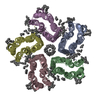 9r21C  9r23C M: map data used to model this data C: citing same article ( |
|---|---|
| Similar structure data |


-Links
 PDBj
PDBj


- Assembly
| Deposited unit | 
|
|---|---|
| 1 |
|
-Components
| #1: Protein | Mass: 26467.082 Da / Num. of mol.: 5 Source method: isolated from a genetically manipulated source Source: (gene. exp.) Candidatus Pseudothioglobus sp. (bacteria)Production host: #2: Chemical | ChemComp-LFA /   Mass: 282.547 Da / Num. of mol.: 30 / Source method: obtained synthetically / Formula: C20H42 Mass: 282.547 Da / Num. of mol.: 30 / Source method: obtained synthetically / Formula: C20H42#3: Chemical | ChemComp-RET /   Mass: 284.436 Da / Num. of mol.: 5 Mass: 284.436 Da / Num. of mol.: 5Source method: isolated from a genetically manipulated source Formula: C20H28O Source: (gene. exp.) Candidatus Pseudothioglobus sp. (bacteria)Production host: #4: Sugar | ChemComp-LMT /   Type: D-saccharide / Mass: 510.615 Da / Num. of mol.: 5 / Source method: obtained synthetically / Formula: C24H46O11 / Comment: detergent*YM Type: D-saccharide / Mass: 510.615 Da / Num. of mol.: 5 / Source method: obtained synthetically / Formula: C24H46O11 / Comment: detergent*YM#5: Water | ChemComp-HOH / |  Mass: 18.015 Da / Num. of mol.: 105 / Source method: isolated from a natural source / Formula: H2O Mass: 18.015 Da / Num. of mol.: 105 / Source method: isolated from a natural source / Formula: H2OHas ligand of interest | Y | Has protein modification | Y | |
|---|
-Experimental details
-Experiment
| Experiment | Method: ELECTRON MICROSCOPY |
|---|---|
| EM experiment | Aggregation state: PARTICLE / 3D reconstruction method: single particle reconstruction |
- Sample preparation
| Component | Name: proteorhodopsin / Type: COMPLEX / Entity ID: #1 / Source: RECOMBINANT |
|---|---|
| Molecular weight | Value: 0.15 MDa / Experimental value: NO |
| Source (natural) | Organism: Candidatus Pseudothioglobus sp. (bacteria) |
| Source (recombinant) | Organism: |
| Buffer solution | pH: 8 |
| Specimen | Conc.: 8 mg/ml / Embedding applied: NO / Shadowing applied: NO / Staining applied: NO / Vitrification applied: YES |
| Specimen support | Details: 5 mA / Grid material: GOLD / Grid mesh size: 300 divisions/in. / Grid type: Quantifoil R1.2/1.3 |
| Vitrification | Instrument: FEI VITROBOT MARK IV / Cryogen name: ETHANE-PROPANE / Humidity: 100 % / Chamber temperature: 288 K / Details: Blotting time 4-6 second, force 0 |
- Electron microscopy imaging
| Experimental equipment |  Model: Titan Krios / Image courtesy: FEI Company |
|---|---|
| Microscopy | Model: TFS KRIOS |
| Electron gun | Electron source:  FIELD EMISSION GUN / Accelerating voltage: 300 kV / Illumination mode: FLOOD BEAM FIELD EMISSION GUN / Accelerating voltage: 300 kV / Illumination mode: FLOOD BEAM |
| Electron lens | Mode: BRIGHT FIELD / Nominal defocus max: 2000 nm / Nominal defocus min: 500 nm |
| Image recording | Electron dose: 50 e/Å2 / Film or detector model: GATAN K3 BIOQUANTUM (6k x 4k) |
- Processing
| CTF correction | Type: PHASE FLIPPING AND AMPLITUDE CORRECTION | ||||||||||||||||||||||||||||||||||||||||||||||||||||||||||||||||||||||||||||||||||||||||||||||||||||||||||
|---|---|---|---|---|---|---|---|---|---|---|---|---|---|---|---|---|---|---|---|---|---|---|---|---|---|---|---|---|---|---|---|---|---|---|---|---|---|---|---|---|---|---|---|---|---|---|---|---|---|---|---|---|---|---|---|---|---|---|---|---|---|---|---|---|---|---|---|---|---|---|---|---|---|---|---|---|---|---|---|---|---|---|---|---|---|---|---|---|---|---|---|---|---|---|---|---|---|---|---|---|---|---|---|---|---|---|---|
| 3D reconstruction | Resolution: 2.48 Å / Resolution method: FSC 0.143 CUT-OFF / Num. of particles: 497291 / Symmetry type: POINT | ||||||||||||||||||||||||||||||||||||||||||||||||||||||||||||||||||||||||||||||||||||||||||||||||||||||||||
| Refinement | Resolution: 2.48→105.34 Å / Cor.coef. Fo:Fc: 0.601 / SU B: 5.579 / SU ML: 0.113 / ESU R: 0.333 Stereochemistry target values: MAXIMUM LIKELIHOOD WITH PHASES Details: HYDROGENS HAVE BEEN USED IF PRESENT IN THE INPUT
| ||||||||||||||||||||||||||||||||||||||||||||||||||||||||||||||||||||||||||||||||||||||||||||||||||||||||||
| Solvent computation | Solvent model: PARAMETERS FOR MASK CACLULATION | ||||||||||||||||||||||||||||||||||||||||||||||||||||||||||||||||||||||||||||||||||||||||||||||||||||||||||
| Displacement parameters | Biso mean: 44.139 Å2 | ||||||||||||||||||||||||||||||||||||||||||||||||||||||||||||||||||||||||||||||||||||||||||||||||||||||||||
| Refinement step | Cycle: 1 / Total: 9643 | ||||||||||||||||||||||||||||||||||||||||||||||||||||||||||||||||||||||||||||||||||||||||||||||||||||||||||
| Refine LS restraints |
|