 Movie
Movie Controller

[English] 日本語
 Yorodumi
- EMDB-53520: Cryo-EM structure of the light-driven proton pump PsPR in deterge... -
+ Open data

- Basic information
| Entry |  | ||||||||||||
|---|---|---|---|---|---|---|---|---|---|---|---|---|---|
| Title | Cryo-EM structure of the light-driven proton pump PsPR in detergent micelle | ||||||||||||
 Map data Map data | Sharpened map used for manual and automatic model refinement | ||||||||||||
 Sample Sample |
| ||||||||||||
 Keywords Keywords | proton transport / retinal / bioenergetics / proteorhodopsin / microbial rhodopsin / MEMBRANE PROTEIN | ||||||||||||
| Biological species |  Candidatus Pseudothioglobus sp. (bacteria) Candidatus Pseudothioglobus sp. (bacteria) | ||||||||||||
| Method | single particle reconstruction / cryo EM / Resolution: 2.48 Å | ||||||||||||
 Authors Authors | Kovalev K / Stetsenko A / Guskov A | ||||||||||||
| Funding support |  Netherlands, Netherlands,  Germany, 3 items Germany, 3 items
| ||||||||||||
 Citation Citation | Journal: Structure / Year: 2025 Title: Structural basis for no retinal binding in flotillin-associated rhodopsins. Authors: Kirill Kovalev / Artem Stetsenko / Florian Trunk / Egor Marin / Jose M Haro-Moreno / Gerrit H U Lamm / Alexey Alekseev / Francisco Rodriguez-Valera / Thomas R Schneider / Josef Wachtveitl / Albert Guskov /  Abstract: Rhodopsins are light-sensitive membrane proteins capturing solar energy via a retinal cofactor covalently attached to a lysine residue. Several groups of rhodopsins lack the conserved lysine and ...Rhodopsins are light-sensitive membrane proteins capturing solar energy via a retinal cofactor covalently attached to a lysine residue. Several groups of rhodopsins lack the conserved lysine and showed no retinal binding. Recently, flotillin-associated rhodopsins (FArhodopsins or FARs) were identified and suggested to lack the retinal-binding pocket despite preserving the lysine residue in many members of the group. Here, we present cryoelectron microscopic (cryo-EM) structures of paralog FArhodopsin and proteorhodopsin from marine bacterium Pseudothioglobus, both forming pentamers similar to those of other microbial rhodopsins. We demonstrate no binding of retinal to the FArhodopsin despite preservation of the lysine residue and overall similarity of the protein fold and internal organization to those of the retinal-binding paralog. Mutational analysis confirmed that two amino acids, H84 and E120, prevent retinal binding within the FArhodopsin. Our work provides insights into the natural retinal loss in microbial rhodopsins and might contribute to the further understanding of FArhodopsins. | ||||||||||||
| History |
|
- Structure visualization
| Supplemental images |
|---|

- Downloads & links
-EMDB archive
| Map data | emd_53520.map.gz | 78.7 MB |  EMDB map data format EMDB map data format | |
|---|---|---|---|---|
| Header (meta data) | emd-53520-v30.xmlemd-53520.xml | 20.2 KB 20.2 KB | Display Display | EMDB header |
| Images |  emd_53520.png emd_53520.png | 68 KB | ||
| Filedesc metadata | emd-53520.cif.gz | 6.1 KB | ||
| Others | emd_53520_additional_1.map.gzemd_53520_half_map_1.map.gzemd_53520_half_map_2.map.gz | 40.5 MB 77.5 MB 77.5 MB | ||
| Archive directory |  http://ftp.pdbj.org/pub/emdb/structures/EMD-53520ftp://ftp.pdbj.org/pub/emdb/structures/EMD-53520 http://ftp.pdbj.org/pub/emdb/structures/EMD-53520ftp://ftp.pdbj.org/pub/emdb/structures/EMD-53520 | HTTPS FTP |
-Related structure data
| Related structure data |  9r22MC 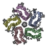 9r21C  9r23C M: atomic model generated by this map C: citing same article ( |
|---|


-Links
| EMDB pages | EMDB (EBI/PDBe) / EMDataResource |
|---|
-Map
| File | Download / File: emd_53520.map.gz / Format: CCP4 / Size: 83.7 MB / Type: IMAGE STORED AS FLOATING POINT NUMBER (4 BYTES) | ||||||||||||||||||||||||||||||||||||
|---|---|---|---|---|---|---|---|---|---|---|---|---|---|---|---|---|---|---|---|---|---|---|---|---|---|---|---|---|---|---|---|---|---|---|---|---|---|
| Annotation | Sharpened map used for manual and automatic model refinement | ||||||||||||||||||||||||||||||||||||
| Projections & slices | Image control
Images are generated by Spider. | ||||||||||||||||||||||||||||||||||||
| Voxel size | X=Y=Z: 0.836 Å | ||||||||||||||||||||||||||||||||||||
| Density |
| ||||||||||||||||||||||||||||||||||||
| Symmetry | Space group: 1 | ||||||||||||||||||||||||||||||||||||
| Details | EMDB XML:
|
 Z (Sec.)
 Y (Row.)
 X (Col.)


-Supplemental data
-Additional map: Non-sharpened map used for validation against coordinates
| File | emd_53520_additional_1.map | ||||||||||||
|---|---|---|---|---|---|---|---|---|---|---|---|---|---|
| Annotation | Non-sharpened map used for validation against coordinates | ||||||||||||
| Projections & Slices |
| ||||||||||||
| Density Histograms |


-Half map: Half map A
| File | emd_53520_half_map_1.map | ||||||||||||
|---|---|---|---|---|---|---|---|---|---|---|---|---|---|
| Annotation | Half map A | ||||||||||||
| Projections & Slices |
| ||||||||||||
| Density Histograms |


-Half map: Half map B
| File | emd_53520_half_map_2.map | ||||||||||||
|---|---|---|---|---|---|---|---|---|---|---|---|---|---|
| Annotation | Half map B | ||||||||||||
| Projections & Slices |
| ||||||||||||
| Density Histograms |


- Sample components
-Entire : proteorhodopsin
| Entire | Name: proteorhodopsin |
|---|---|
| Components |
|
-Supramolecule #1: proteorhodopsin
| Supramolecule | Name: proteorhodopsin / type: complex / ID: 1 / Parent: 0 / Macromolecule list: #1 |
|---|---|
| Source (natural) | Organism: Candidatus Pseudothioglobus sp. (bacteria) |
| Molecular weight | Theoretical: 150 KDa |
-Macromolecule #1: microbial rhodopsin
| Macromolecule | Name: microbial rhodopsin / type: protein_or_peptide / ID: 1 / Number of copies: 5 / Enantiomer: LEVO |
|---|---|
| Source (natural) | Organism: Candidatus Pseudothioglobus sp. (bacteria) |
| Molecular weight | Theoretical: 26.467082 KDa |
| Recombinant expression | Organism: |
| Sequence | String: MLQAGDFVGV SFWLVSVAMV AATVFFFYEG MSVKKEWKLS MTIAGLVTLV AAIHYYYMRD YWVASVLAGS PDSPIVYRYI DWLITVPLL MIEFFIILKA VGASISTNSF WRLLVGTLVM LIGGFAGEAM LISASLGFII GMVGWAIIIW EIFGGEASKA A DANAGVKS ...String: MLQAGDFVGV SFWLVSVAMV AATVFFFYEG MSVKKEWKLS MTIAGLVTLV AAIHYYYMRD YWVASVLAGS PDSPIVYRYI DWLITVPLL MIEFFIILKA VGASISTNSF WRLLVGTLVM LIGGFAGEAM LISASLGFII GMVGWAIIIW EIFGGEASKA A DANAGVKS AFNALRLIVL VGWAIYPLGY IFGYMMGSVD SGSLNIIYNL ADFVNKILFG LIIWNVAVRE SSDALEHHHH HH |
-Macromolecule #2: EICOSANE
| Macromolecule | Name: EICOSANE / type: ligand / ID: 2 / Number of copies: 30 / Formula: LFA |
|---|---|
| Molecular weight | Theoretical: 282.547 Da |
| Chemical component information |  ChemComp-LFA: |
-Macromolecule #3: RETINAL
| Macromolecule | Name: RETINAL / type: ligand / ID: 3 / Number of copies: 5 / Formula: RET |
|---|---|
| Source (natural) | Organism: Candidatus Pseudothioglobus sp. (bacteria) |
| Molecular weight | Theoretical: 284.436 Da |
| Chemical component information |  ChemComp-RET: |
-Macromolecule #4: DODECYL-BETA-D-MALTOSIDE
| Macromolecule | Name: DODECYL-BETA-D-MALTOSIDE / type: ligand / ID: 4 / Number of copies: 5 / Formula: LMT |
|---|---|
| Molecular weight | Theoretical: 510.615 Da |
| Chemical component information |  ChemComp-LMT: |
-Macromolecule #5: water
| Macromolecule | Name: water / type: ligand / ID: 5 / Number of copies: 105 / Formula: HOH |
|---|---|
| Molecular weight | Theoretical: 18.015 Da |
| Chemical component information |  ChemComp-HOH: |
-Experimental details
-Structure determination
| Method | cryo EM |
|---|---|
 Processing Processing | single particle reconstruction |
| Aggregation state | particle |
-Sample preparation
| Concentration | 8 mg/mL |
|---|---|
| Buffer | pH: 8 |
| Grid | Model: Quantifoil R1.2/1.3 / Material: GOLD / Mesh: 300 / Pretreatment - Type: GLOW DISCHARGE / Pretreatment - Time: 30 sec. / Details: 5 mA |
| Vitrification | Cryogen name: ETHANE-PROPANE / Chamber humidity: 100 % / Chamber temperature: 288 K / Instrument: FEI VITROBOT MARK IV / Details: Blotting time 4-6 second, force 0. |
- Electron microscopy
| Microscope | TFS KRIOS |
|---|---|
| Image recording | Film or detector model: GATAN K3 BIOQUANTUM (6k x 4k) / Average electron dose: 50.0 e/Å2 |
| Electron beam | Acceleration voltage: 300 kV / Electron source:  FIELD EMISSION GUN FIELD EMISSION GUN |
| Electron optics | Illumination mode: FLOOD BEAM / Imaging mode: BRIGHT FIELD / Nominal defocus max: 2.0 µm / Nominal defocus min: 0.5 µm |
| Experimental equipment |  Model: Titan Krios / Image courtesy: FEI Company |
