 Movie
Movie Controller

[English] 日本語
 Yorodumi
- PDB-9bdc: Cryo-EM Structure of the TEFM-bound Human Mitochondrial Transcrip... -
+ Open data

- Basic information
| Entry | Database: PDB / ID: 9bdc | ||||||
|---|---|---|---|---|---|---|---|
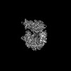
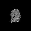
| Title | Cryo-EM Structure of the TEFM-bound Human Mitochondrial Transcription Substrate Rejection Complex | ||||||
 Components Components |
| ||||||
 Keywords Keywords | TRANSCRIPTION/DNA/RNA / Mitochondrial RNA Polymerase / Nucleotide Selection / Nucleotide Discrimination / TRANSCRIPTION / Protein-RNA-DNA Complex / POLRMT / TEFM / TRANSCRIPTION-DNA-RNA complex | ||||||
| Function / homology |  Function and homology information Function and homology informationtranscription elongation by mitochondrial RNA polymerase / positive regulation of mitochondrial transcription / Mitochondrial transcription initiation / mitochondrial DNA-directed RNA polymerase complex / mitochondrial promoter sequence-specific DNA binding / transcription initiation at mitochondrial promoter / regulation of oxidative phosphorylation / Strand-asynchronous mitochondrial DNA replication / mitochondrial DNA replication / mitochondrial transcription ...transcription elongation by mitochondrial RNA polymerase / positive regulation of mitochondrial transcription / Mitochondrial transcription initiation / mitochondrial DNA-directed RNA polymerase complex / mitochondrial promoter sequence-specific DNA binding / transcription initiation at mitochondrial promoter / regulation of oxidative phosphorylation / Strand-asynchronous mitochondrial DNA replication / mitochondrial DNA replication / mitochondrial transcription / transcription elongation factor activity / mitochondrial nucleoid / Transcriptional activation of mitochondrial biogenesis / DNA-directed RNA polymerase / DNA-directed RNA polymerase activity / 3'-5'-RNA exonuclease activity / sequence-specific DNA binding / mitochondrial matrix / ribonucleoprotein complex / protein-containing complex / mitochondrion / RNA binding Similarity search - Function | ||||||
| Biological species |  Homo sapiens (human) Homo sapiens (human)synthetic construct (others) | ||||||
| Method | ELECTRON MICROSCOPY / single particle reconstruction / cryo EM / Resolution: 2.54 Å | ||||||
 Authors Authors | Herbine, K.H. / Nayak, A.R. / Temiakov, D. | ||||||
| Funding support |  United States, 1items United States, 1items
| ||||||
 Citation Citation | Journal: Nat Commun / Year: 2024 Title: Structural basis for substrate binding and selection by human mitochondrial RNA polymerase. Authors: Karl Herbine / Ashok R Nayak / Dmitry Temiakov / Abstract: The mechanism by which RNAP selects cognate substrates and discriminates between deoxy and ribonucleotides is of fundamental importance to the fidelity of transcription. Here, we present cryo-EM ...The mechanism by which RNAP selects cognate substrates and discriminates between deoxy and ribonucleotides is of fundamental importance to the fidelity of transcription. Here, we present cryo-EM structures of human mitochondrial transcription elongation complexes that reveal substrate ATP bound in Entry and Insertion Sites. In the Entry Site, the substrate binds along the O helix of the fingers domain of mtRNAP but does not interact with the templating DNA base. Interactions between RNAP and the triphosphate moiety of the NTP in the Entry Site ensure discrimination against nucleosides and their diphosphate and monophosphate derivatives but not against non-cognate rNTPs and dNTPs. Closing of the fingers domain over the catalytic site results in delivery of both the templating DNA base and the substrate into the Insertion Site and recruitment of the catalytic magnesium ions. The cryo-EM data also reveal a conformation adopted by mtRNAP to reject a non-cognate substrate from its active site. Our findings establish a structural basis for substrate binding and suggest a unified mechanism of NTP selection for single-subunit RNAPs. | ||||||
| History |
|
- Structure visualization
| Structure viewer | Molecule: MolmilJmol/JSmol |
|---|
- Downloads & links
-Download
| PDBx/mmCIF format | 9bdc.cif.gz | 324.4 KB | Display | PDBx/mmCIF format |
|---|---|---|---|---|
| PDB format | pdb9bdc.ent.gz | 242.7 KB | Display | PDB format |
| PDBx/mmJSON format | 9bdc.json.gz | Tree view | PDBx/mmJSON format | |
| Others |  Other downloads Other downloads |
-Validation report
| Arichive directory | https://data.pdbj.org/pub/pdb/validation_reports/bd/9bdcftp://data.pdbj.org/pub/pdb/validation_reports/bd/9bdc | HTTPS FTP |
|---|
-Related structure data
| Related structure data |  44448MC 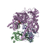 8u8uC  8u8vC 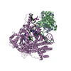 9bddC M: map data used to model this data C: citing same article ( |
|---|---|
| Similar structure data |
-Links
 PDBj
PDBj


- Assembly
| Deposited unit | 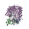
|
|---|---|
| 1 |
|
-Components
| #1: Protein | Mass: 27262.389 Da / Num. of mol.: 2 Source method: isolated from a genetically manipulated source Source: (gene. exp.) Homo sapiens (human) / Gene: TEFM, C17orf42 / Production host:  #2: Protein | | Mass: 127150.102 Da / Num. of mol.: 1 Source method: isolated from a genetically manipulated source Source: (gene. exp.) Homo sapiens (human) / Gene: POLRMT / Production host: #3: DNA chain | | Mass: 10474.738 Da / Num. of mol.: 1 / Source method: obtained synthetically / Source: (synth.) synthetic construct (others) #4: RNA chain | | Mass: 4493.723 Da / Num. of mol.: 1 / Source method: obtained synthetically / Source: (synth.) synthetic construct (others) #5: DNA chain | | Mass: 10428.664 Da / Num. of mol.: 1 / Source method: obtained synthetically / Source: (synth.) synthetic construct (others) Has protein modification | N | |
|---|
-Experimental details
-Experiment
| Experiment | Method: ELECTRON MICROSCOPY |
|---|---|
| EM experiment | Aggregation state: PARTICLE / 3D reconstruction method: single particle reconstruction |
- Sample preparation
| Component | Name: Structure of the Transcription Elongation Complex of Human Mitochondrial Polymerase with the Closed Conformation of the Fingers Domain Type: COMPLEX Details: Human Mitochondrial RNA polymerase (d119) and Human TEFM (d135) assembled on an RNA-DNA Scaffold in presence of methylene a,b-ATP Entity ID: all / Source: RECOMBINANT |
|---|---|
| Molecular weight | Value: 0.227 MDa / Experimental value: NO |
| Source (natural) | Organism: Homo sapiens (human) |
| Source (recombinant) | Organism: |
| Buffer solution | pH: 7.9 Details: 20 mM Tris Buffer, pH 7.9, 150 mM NaCl, 10 mM DTT, and 5 mM MgCl2 |
| Specimen | Conc.: 0.75 mg/ml / Embedding applied: NO / Shadowing applied: NO / Staining applied: NO / Vitrification applied: YES Details: 5 uM complex of (D119) wild-type mtRNAP, (D50) wild-type TEFM, an RNA-DNA scaffold (R14/T31_+1A/NT27_+1T), and a,b methylene ATP. |
| Specimen support | Details: 15 mA Discharge / Grid material: GOLD / Grid mesh size: 300 divisions/in. / Grid type: UltrAuFoil R1.2/1.3 |
| Vitrification | Instrument: FEI VITROBOT MARK IV / Cryogen name: ETHANE / Humidity: 100 % / Chamber temperature: 277 K |
- Electron microscopy imaging
| Experimental equipment |  Model: Titan Krios / Image courtesy: FEI Company |
|---|---|
| Microscopy | Model: FEI TITAN KRIOS Details: Preliminary grid screening performed manually using TFS Glacios. |
| Electron gun | Electron source:  FIELD EMISSION GUN / Accelerating voltage: 300 kV / Illumination mode: FLOOD BEAM FIELD EMISSION GUN / Accelerating voltage: 300 kV / Illumination mode: FLOOD BEAM |
| Electron lens | Mode: BRIGHT FIELD / Nominal magnification: 105000 X / Nominal defocus max: 1800 nm / Nominal defocus min: 500 nm / Cs: 2.7 mm / C2 aperture diameter: 100 µm / Alignment procedure: COMA FREE |
| Specimen holder | Cryogen: NITROGEN / Specimen holder model: FEI TITAN KRIOS AUTOGRID HOLDER |
| Image recording | Average exposure time: 1.89 sec. / Electron dose: 50.6 e/Å2 / Film or detector model: GATAN K3 (6k x 4k) / Num. of grids imaged: 2 / Num. of real images: 21764 |
| EM imaging optics | Energyfilter slit width: 20 eV |
- Processing
| EM software | Name: PHENIX / Version: 1.21rc1_5015: / Category: model refinement | ||||||||||||||||||||||||
|---|---|---|---|---|---|---|---|---|---|---|---|---|---|---|---|---|---|---|---|---|---|---|---|---|---|
| CTF correction | Type: PHASE FLIPPING AND AMPLITUDE CORRECTION | ||||||||||||||||||||||||
| Particle selection | Num. of particles selected: 7998459 Details: Automated particle picking (Blob picker, CryoSPARC) with manual inspection (Inspect Particle Picks). | ||||||||||||||||||||||||
| Symmetry | Point symmetry: C1 (asymmetric) | ||||||||||||||||||||||||
| 3D reconstruction | Resolution: 2.54 Å / Resolution method: FSC 0.143 CUT-OFF / Num. of particles: 2457845 / Algorithm: BACK PROJECTION Details: Non-uniform Refinement (CryoSPARC) which is an algorithm based on cross-validation optimization, which automatically regularizes 3D density maps during refinement to account for spatial variability. Num. of class averages: 1 / Symmetry type: POINT | ||||||||||||||||||||||||
| Atomic model building | B value: 45 / Protocol: FLEXIBLE FIT / Space: REAL / Target criteria: Cross-correlation Coefficient Details: Initial Fitting was done in Chimera and flexible/refined fitting done with Coot Phenix Real-Space Refinement. | ||||||||||||||||||||||||
| Atomic model building | PDB-ID: 5OLA Accession code: 5OLA / Source name: PDB / Type: experimental model | ||||||||||||||||||||||||
| Refine LS restraints |
|

