 Movie
Movie Controller

[English] 日本語
 Yorodumi
- PDB-8y65: Cryo-EM structure of human urate transporter GLUT9 bound to subst... -
+ Open data

- Basic information
| Entry | Database: PDB / ID: 8y65 | |||||||||
|---|---|---|---|---|---|---|---|---|---|---|
| Title | Cryo-EM structure of human urate transporter GLUT9 bound to substrate urate | |||||||||
 Components Components | Solute carrier family 2, facilitated glucose transporter member 9 | |||||||||
 Keywords Keywords | TRANSPORT PROTEIN / GLUT9 / Urate | |||||||||
| Function / homology |  Function and homology information Function and homology informationDefective SLC2A9 causes hypouricemia renal 2 (RHUC2) / fructose transmembrane transporter activity / fructose transmembrane transport / dehydroascorbic acid transport / hexose transmembrane transport / carbohydrate:proton symporter activity / Cellular hexose transport / D-glucose transmembrane transporter activity / D-glucose transmembrane transport / urate transport ...Defective SLC2A9 causes hypouricemia renal 2 (RHUC2) / fructose transmembrane transporter activity / fructose transmembrane transport / dehydroascorbic acid transport / hexose transmembrane transport / carbohydrate:proton symporter activity / Cellular hexose transport / D-glucose transmembrane transporter activity / D-glucose transmembrane transport / urate transport / urate metabolic process / urate transmembrane transporter activity / : / transmembrane transporter activity / basolateral plasma membrane / apical plasma membrane / membrane / plasma membrane Similarity search - Function | |||||||||
| Biological species |  Homo sapiens (human) Homo sapiens (human) | |||||||||
| Method | ELECTRON MICROSCOPY / single particle reconstruction / cryo EM / Resolution: 3.51 Å | |||||||||
 Authors Authors | Pan, X.J. / Shen, Z.L. / Xu, L. / Huang, G.X.Y. | |||||||||
| Funding support |  China, 2items China, 2items
| |||||||||
 Citation Citation | Journal: Nat Commun / Year: 2024 Title: Structural basis for urate recognition and apigenin inhibition of human GLUT9. Authors: Zilin Shen / Li Xu / Tong Wu / Huan Wang / Qifan Wang / Xiaofei Ge / Fang Kong / Gaoxingyu Huang / Xiaojing Pan / Abstract: Urate, the physiological form of uric acid and a potent antioxidant in serum, plays a pivotal role in scavenging reactive oxygen species. Yet excessive accumulation of urate, known as hyperuricemia, ...Urate, the physiological form of uric acid and a potent antioxidant in serum, plays a pivotal role in scavenging reactive oxygen species. Yet excessive accumulation of urate, known as hyperuricemia, is the primary risk factor for the development of gout. The high-capacity urate transporter GLUT9 represents a promising target for gout treatment. Here, we present cryo-electron microscopy structures of human GLUT9 in complex with urate or its inhibitor apigenin at overall resolutions of 3.5 Å and 3.3 Å, respectively. In both structures, GLUT9 exhibits an inward open conformation, wherein the substrate binding pocket faces the intracellular side. These structures unveil the molecular basis for GLUT9's substrate preference of urate over glucose, and show that apigenin acts as a competitive inhibitor by occupying the substrate binding site. Our findings provide critical information for the development of specific inhibitors targeting GLUT9 as potential therapeutics for gout and hyperuricemia. | |||||||||
| History |
|
- Structure visualization
| Structure viewer | Molecule: MolmilJmol/JSmol |
|---|
- Downloads & links
-Download
| PDBx/mmCIF format | 8y65.cif.gz | 93 KB | Display | PDBx/mmCIF format |
|---|---|---|---|---|
| PDB format | pdb8y65.ent.gz | 67.3 KB | Display | PDB format |
| PDBx/mmJSON format | 8y65.json.gz | Tree view | PDBx/mmJSON format | |
| Others |  Other downloads Other downloads |
-Validation report
| Arichive directory | https://data.pdbj.org/pub/pdb/validation_reports/y6/8y65ftp://data.pdbj.org/pub/pdb/validation_reports/y6/8y65 | HTTPS FTP |
|---|
-Related structure data
| Related structure data |  38966MC 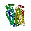 8y66C M: map data used to model this data C: citing same article ( |
|---|---|
| Similar structure data |
-Links
 PDBj
PDBj


- Assembly
| Deposited unit | 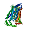
|
|---|---|
| 1 |
|
-Components
| #1: Protein | Mass: 63215.090 Da / Num. of mol.: 1 Source method: isolated from a genetically manipulated source Source: (gene. exp.) Homo sapiens (human) / Gene: SLC2A9, GLUT9 / Production host: Homo sapiens (human) / References: UniProt: Q9NRM0 |
|---|---|
| #2: Chemical | ChemComp-URC /   Mass: 168.110 Da / Num. of mol.: 1 / Source method: obtained synthetically / Formula: C5H4N4O3 / Feature type: SUBJECT OF INVESTIGATION Mass: 168.110 Da / Num. of mol.: 1 / Source method: obtained synthetically / Formula: C5H4N4O3 / Feature type: SUBJECT OF INVESTIGATION |
| Has ligand of interest | Y |
-Experimental details
-Experiment
| Experiment | Method: ELECTRON MICROSCOPY |
|---|---|
| EM experiment | Aggregation state: PARTICLE / 3D reconstruction method: single particle reconstruction |
- Sample preparation
| Component | Name: GLUT9 in complex with urate / Type: COMPLEX / Entity ID: #1 / Source: RECOMBINANT |
|---|---|
| Source (natural) | Organism: Homo sapiens (human) |
| Source (recombinant) | Organism: Homo sapiens (human) |
| Buffer solution | pH: 6 |
| Specimen | Conc.: 1 mg/ml / Embedding applied: NO / Shadowing applied: NO / Staining applied: NO / Vitrification applied: YES |
| Vitrification | Cryogen name: ETHANE / Humidity: 100 % |
- Electron microscopy imaging
| Experimental equipment |  Model: Titan Krios / Image courtesy: FEI Company |
|---|---|
| Microscopy | Model: FEI TITAN KRIOS |
| Electron gun | Electron source:  FIELD EMISSION GUN / Accelerating voltage: 300 kV / Illumination mode: FLOOD BEAM FIELD EMISSION GUN / Accelerating voltage: 300 kV / Illumination mode: FLOOD BEAM |
| Electron lens | Mode: BRIGHT FIELD / Nominal defocus max: 1800 nm / Nominal defocus min: 1500 nm |
| Image recording | Average exposure time: 2.56 sec. / Electron dose: 50 e/Å2 / Film or detector model: GATAN K3 (6k x 4k) |
- Processing
| EM software | Name: PHENIX / Version: 1.19.2_4158: / Category: model refinement | ||||||||||||||||||||||||
|---|---|---|---|---|---|---|---|---|---|---|---|---|---|---|---|---|---|---|---|---|---|---|---|---|---|
| CTF correction | Type: PHASE FLIPPING AND AMPLITUDE CORRECTION | ||||||||||||||||||||||||
| 3D reconstruction | Resolution: 3.51 Å / Resolution method: FSC 0.143 CUT-OFF / Num. of particles: 810830 / Symmetry type: POINT | ||||||||||||||||||||||||
| Refine LS restraints |
|
