 Movie
Movie Controller


+ Open data

- Basic information
| Entry | Database: PDB / ID: 8xvb | |||||||||||||||||||||||||||||||||||||||
|---|---|---|---|---|---|---|---|---|---|---|---|---|---|---|---|---|---|---|---|---|---|---|---|---|---|---|---|---|---|---|---|---|---|---|---|---|---|---|---|---|
| Title | Cryo-EM structure of ATP-DNA-MuB filaments | |||||||||||||||||||||||||||||||||||||||
 Components Components |
| |||||||||||||||||||||||||||||||||||||||
 Keywords Keywords | VIRAL PROTEIN/DNA / VIRAL PROTEIN-DNA COMPLEX | |||||||||||||||||||||||||||||||||||||||
| Function / homology |  Function and homology information Function and homology informationDNA transposition / viral DNA genome replication / Hydrolases; Acting on acid anhydrides; In phosphorus-containing anhydrides / DNA integration / host cell cytoplasm / DNA replication / ATP hydrolysis activity / DNA binding / ATP binding / metal ion binding Similarity search - Function | |||||||||||||||||||||||||||||||||||||||
| Biological species |  Escherichia phage Mu (virus) Escherichia phage Mu (virus) | |||||||||||||||||||||||||||||||||||||||
| Method | ELECTRON MICROSCOPY / helical reconstruction / cryo EM / Resolution: 3.4 Å | |||||||||||||||||||||||||||||||||||||||
 Authors Authors | Zhao, X. / Zhang, K. / Li, S. | |||||||||||||||||||||||||||||||||||||||
| Funding support | 1items
| |||||||||||||||||||||||||||||||||||||||
 Citation Citation | Journal: Nat Commun / Year: 2024 Title: Elucidating the Architectural dynamics of MuB filaments in bacteriophage Mu DNA transposition. Authors: Xiaolong Zhao / Yongxiang Gao / Qingguo Gong / Kaiming Zhang / Shanshan Li /  Abstract: MuB is a non-specific DNA-binding protein and AAA+ ATPase that significantly influences the DNA transposition process of bacteriophage Mu, especially in target DNA selection for transposition. While ...MuB is a non-specific DNA-binding protein and AAA+ ATPase that significantly influences the DNA transposition process of bacteriophage Mu, especially in target DNA selection for transposition. While studies have established the ATP-dependent formation of MuB filament as pivotal to this process, the high-resolution structure of a full-length MuB protomer and the underlying molecular mechanisms governing its oligomerization remain elusive. Here, we use cryo-EM to obtain a 3.4-Å resolution structure of the ATP(+)-DNA(+)-MuB helical filament, which encapsulates the DNA substrate within its axial channel. The structure categorizes MuB within the initiator clade of the AAA+ protein family and precisely locates the ATP and DNA binding sites. Further investigation into the oligomeric states of MuB show the existence of various forms of the filament. These findings lead to a mechanistic model where MuB forms opposite helical filaments along the DNA, exposing potential target sites on the bare DNA and then recruiting MuA, which stimulates MuB's ATPase activity and disrupts the previously formed helical structure. When this happens, MuB generates larger ring structures and dissociates from the DNA. | |||||||||||||||||||||||||||||||||||||||
| History |
|
- Structure visualization
| Structure viewer | Molecule: MolmilJmol/JSmol |
|---|
- Downloads & links
-Download
| PDBx/mmCIF format | 8xvb.cif.gz | 367.7 KB | Display | PDBx/mmCIF format |
|---|---|---|---|---|
| PDB format | pdb8xvb.ent.gz | 300.5 KB | Display | PDB format |
| PDBx/mmJSON format | 8xvb.json.gz | Tree view | PDBx/mmJSON format | |
| Others |  Other downloads Other downloads |
-Validation report
| Arichive directory | https://data.pdbj.org/pub/pdb/validation_reports/xv/8xvbftp://data.pdbj.org/pub/pdb/validation_reports/xv/8xvb | HTTPS FTP |
|---|
-Related structure data
| Related structure data |  38695MC 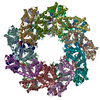 8xvcC  8xvdC M: map data used to model this data C: citing same article ( |
|---|---|
| Similar structure data |


-Links
 PDBj
PDBj


- Assembly
| Deposited unit | 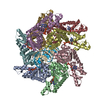
|
|---|---|
| 1 |
|
-Components
| #1: DNA chain | Mass: 7472.007 Da / Num. of mol.: 1 / Source method: obtained synthetically / Source: (synth.) Escherichia phage Mu (virus) | ||||||||
|---|---|---|---|---|---|---|---|---|---|
| #2: Protein | Mass: 35153.133 Da / Num. of mol.: 8 Source method: isolated from a genetically manipulated source Source: (gene. exp.) Escherichia phage Mu (virus) / Gene: B, Mup04Production host:  References: UniProt: P03763, Hydrolases; Acting on acid anhydrides; In phosphorus-containing anhydrides #3: DNA chain | | Mass: 7255.670 Da / Num. of mol.: 1 / Source method: obtained synthetically / Source: (synth.) Escherichia phage Mu (virus)#4: Chemical | ChemComp-ATP /   Mass: 507.181 Da / Num. of mol.: 8 / Source method: obtained synthetically / Formula: C10H16N5O13P3 / Feature type: SUBJECT OF INVESTIGATION / Comment: ATP, energy-carrying molecule*YM Mass: 507.181 Da / Num. of mol.: 8 / Source method: obtained synthetically / Formula: C10H16N5O13P3 / Feature type: SUBJECT OF INVESTIGATION / Comment: ATP, energy-carrying molecule*YMHas ligand of interest | Y | Has protein modification | N | |
-Experimental details
-Experiment
| Experiment | Method: ELECTRON MICROSCOPY |
|---|---|
| EM experiment | Aggregation state: FILAMENT / 3D reconstruction method: helical reconstruction |
- Sample preparation
| Component | Name: ATP-DNA-MuB Filaments in Bacteriophage Mu DNA Transposition Type: COMPLEX / Entity ID: #1-#3 / Source: RECOMBINANT |
|---|---|
| Source (natural) | Organism: Escherichia phage Mu (virus) |
| Source (recombinant) | Organism: |
| Buffer solution | pH: 7.8 |
| Specimen | Embedding applied: NO / Shadowing applied: NO / Staining applied: NO / Vitrification applied: YES |
| Vitrification | Cryogen name: ETHANE |
- Electron microscopy imaging
| Experimental equipment |  Model: Titan Krios / Image courtesy: FEI Company |
|---|---|
| Microscopy | Model: FEI TITAN KRIOS |
| Electron gun | Electron source:  FIELD EMISSION GUN / Accelerating voltage: 300 kV / Illumination mode: FLOOD BEAM FIELD EMISSION GUN / Accelerating voltage: 300 kV / Illumination mode: FLOOD BEAM |
| Electron lens | Mode: BRIGHT FIELD / Nominal defocus max: 3000 nm / Nominal defocus min: 2000 nm |
| Image recording | Electron dose: 60.9 e/Å2 / Film or detector model: GATAN K3 (6k x 4k) / Num. of real images: 6298 |
- Processing
| EM software | Name: PHENIX / Category: model refinement | ||||||||||||||||||||||||
|---|---|---|---|---|---|---|---|---|---|---|---|---|---|---|---|---|---|---|---|---|---|---|---|---|---|
| CTF correction | Type: NONE | ||||||||||||||||||||||||
| Helical symmerty | Angular rotation/subunit: 56.16 ° / Axial rise/subunit: 6.91 Å / Axial symmetry: C1 | ||||||||||||||||||||||||
| 3D reconstruction | Resolution: 3.4 Å / Resolution method: FSC 0.143 CUT-OFF / Num. of particles: 377689 / Symmetry type: HELICAL | ||||||||||||||||||||||||
| Refine LS restraints |
|
