 Movie
Movie Controller


+ Open data

- Basic information
| Entry |  | |||||||||
|---|---|---|---|---|---|---|---|---|---|---|
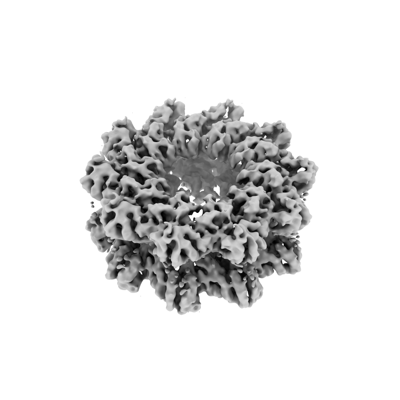
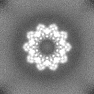
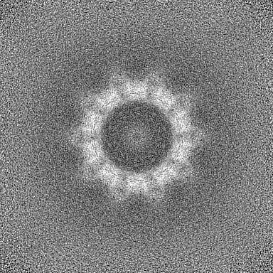
| Title | CryoEM structure of ADP-DNA-MuB conformation2 | |||||||||
 Map data Map data | CryoEM structure of ADP-DNA-MuB conformation2 with D10 | |||||||||
 Sample Sample |
| |||||||||
 Keywords Keywords | ADP-MuB complex / D10 symmetry / Ring / VIRAL PROTEIN | |||||||||
| Function / homology |  Function and homology information Function and homology informationDNA transposition / viral DNA genome replication / Hydrolases; Acting on acid anhydrides; In phosphorus-containing anhydrides / DNA integration / host cell cytoplasm / DNA replication / ATP hydrolysis activity / DNA binding / ATP binding / metal ion binding Similarity search - Function | |||||||||
| Biological species |  Escherichia phage Mu (virus) Escherichia phage Mu (virus) | |||||||||
| Method | single particle reconstruction / cryo EM / Resolution: 4.43 Å | |||||||||
 Authors Authors | Zhao X / Zhang K / Li S | |||||||||
| Funding support | 1 items
| |||||||||
 Citation Citation | Journal: Nat Commun / Year: 2024 Title: Elucidating the Architectural dynamics of MuB filaments in bacteriophage Mu DNA transposition. Authors: Xiaolong Zhao / Yongxiang Gao / Qingguo Gong / Kaiming Zhang / Shanshan Li /  Abstract: MuB is a non-specific DNA-binding protein and AAA+ ATPase that significantly influences the DNA transposition process of bacteriophage Mu, especially in target DNA selection for transposition. While ...MuB is a non-specific DNA-binding protein and AAA+ ATPase that significantly influences the DNA transposition process of bacteriophage Mu, especially in target DNA selection for transposition. While studies have established the ATP-dependent formation of MuB filament as pivotal to this process, the high-resolution structure of a full-length MuB protomer and the underlying molecular mechanisms governing its oligomerization remain elusive. Here, we use cryo-EM to obtain a 3.4-Å resolution structure of the ATP(+)-DNA(+)-MuB helical filament, which encapsulates the DNA substrate within its axial channel. The structure categorizes MuB within the initiator clade of the AAA+ protein family and precisely locates the ATP and DNA binding sites. Further investigation into the oligomeric states of MuB show the existence of various forms of the filament. These findings lead to a mechanistic model where MuB forms opposite helical filaments along the DNA, exposing potential target sites on the bare DNA and then recruiting MuA, which stimulates MuB's ATPase activity and disrupts the previously formed helical structure. When this happens, MuB generates larger ring structures and dissociates from the DNA. | |||||||||
| History |
|
- Structure visualization
| Supplemental images |
|---|
- Downloads & links
-EMDB archive
| Map data | emd_38697.map.gz | 203 MB | EMDB map data format | |
|---|---|---|---|---|
| Header (meta data) | emd-38697-v30.xmlemd-38697.xml | 21.1 KB 21.1 KB | Display Display | EMDB header |
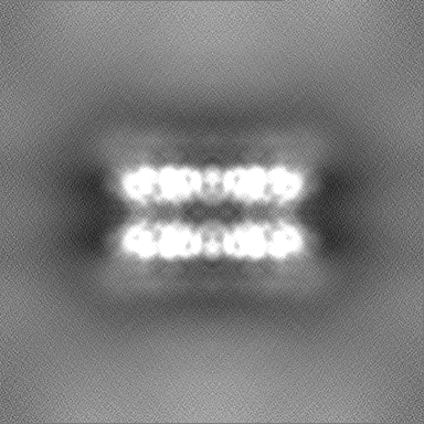
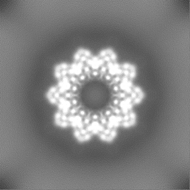
| Images |  emd_38697.png emd_38697.png | 57.5 KB | ||
| Filedesc metadata | emd-38697.cif.gz | 5.8 KB | ||
| Others | emd_38697_additional_1.map.gzemd_38697_additional_2.map.gzemd_38697_half_map_1.map.gzemd_38697_half_map_2.map.gz | 102.8 MB 202.9 MB 200.1 MB 200.1 MB | ||
| Archive directory |  http://ftp.pdbj.org/pub/emdb/structures/EMD-38697ftp://ftp.pdbj.org/pub/emdb/structures/EMD-38697 http://ftp.pdbj.org/pub/emdb/structures/EMD-38697ftp://ftp.pdbj.org/pub/emdb/structures/EMD-38697 | HTTPS FTP |
-Related structure data
| Related structure data |  8xvdMC  8xvbC  8xvcC M: atomic model generated by this map C: citing same article ( |
|---|---|
| Similar structure data |


-Links
| EMDB pages | EMDB (EBI/PDBe) / EMDataResource |
|---|---|
| Related items in Molecule of the Month |


-Map
| File | Download / File: emd_38697.map.gz / Format: CCP4 / Size: 216 MB / Type: IMAGE STORED AS FLOATING POINT NUMBER (4 BYTES) | ||||||||||||||||||||||||||||||||||||
|---|---|---|---|---|---|---|---|---|---|---|---|---|---|---|---|---|---|---|---|---|---|---|---|---|---|---|---|---|---|---|---|---|---|---|---|---|---|
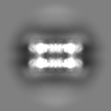
| Annotation | CryoEM structure of ADP-DNA-MuB conformation2 with D10 | ||||||||||||||||||||||||||||||||||||
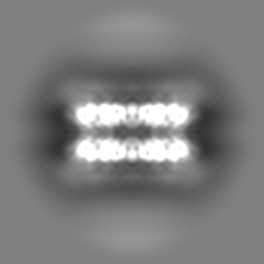
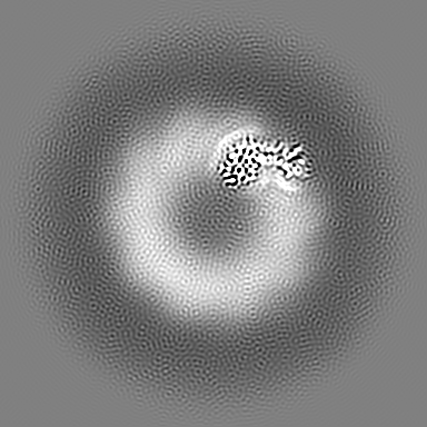
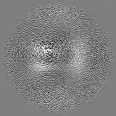
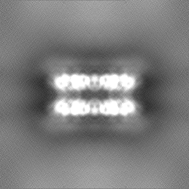
| Projections & slices | Image control
Images are generated by Spider. | ||||||||||||||||||||||||||||||||||||
| Voxel size | X=Y=Z: 0.82 Å | ||||||||||||||||||||||||||||||||||||
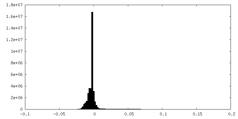
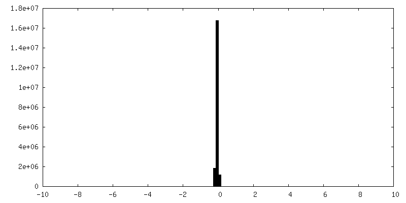
| Density |
| ||||||||||||||||||||||||||||||||||||
| Symmetry | Space group: 1 | ||||||||||||||||||||||||||||||||||||
| Details | EMDB XML:
|
 Z (Sec.)
 Y (Row.)
 X (Col.)


-Supplemental data
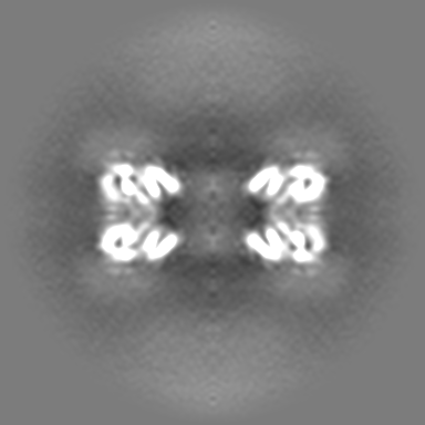
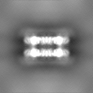
-Additional map: CryoEM structure of ADP-DNA-MuB conformation2 with D10
| File | emd_38697_additional_1.map | ||||||||||||
|---|---|---|---|---|---|---|---|---|---|---|---|---|---|
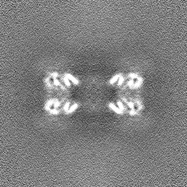
| Annotation | CryoEM structure of ADP-DNA-MuB conformation2 with D10 | ||||||||||||
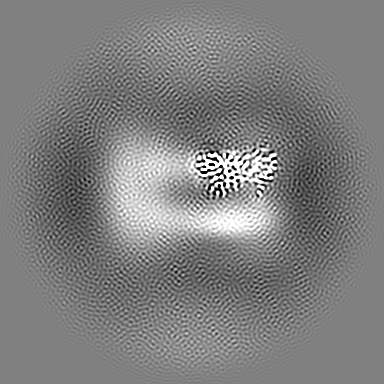
| Projections & Slices |
| ||||||||||||
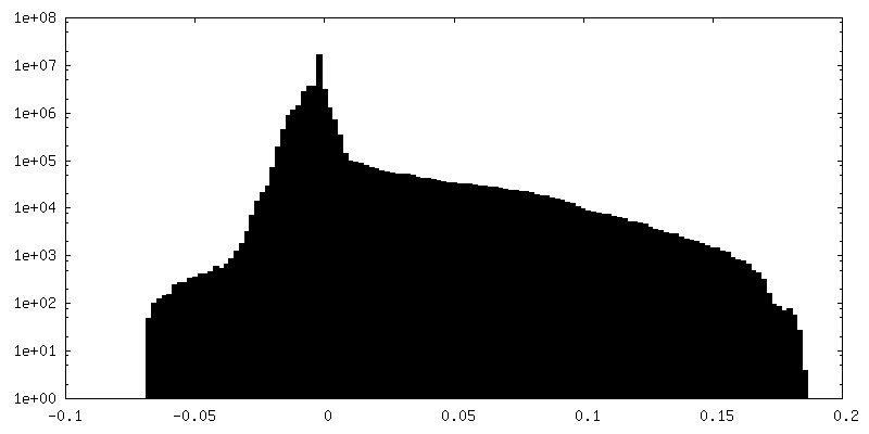
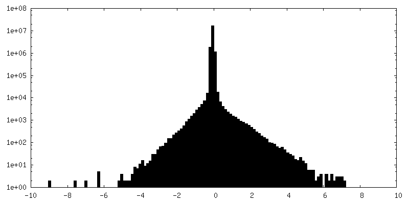
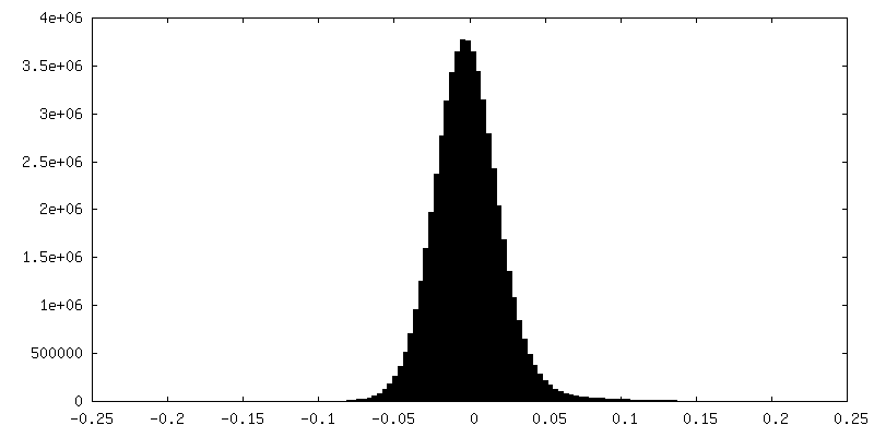
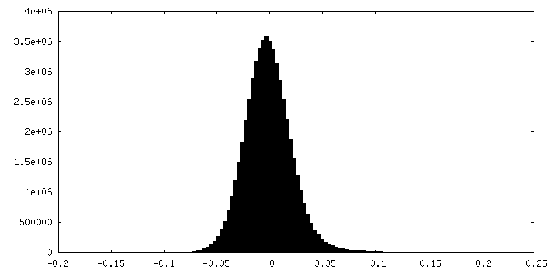
| Density Histograms |


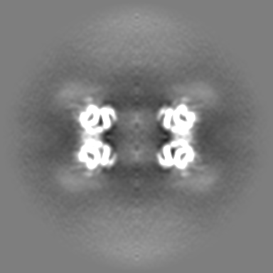
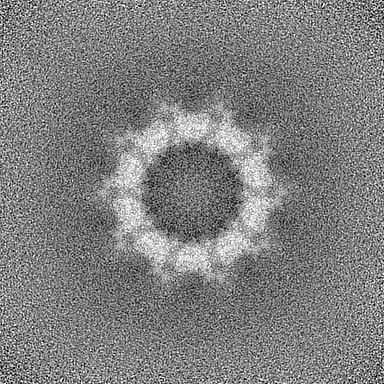
-Additional map: CryoEM structure of ADP-DNA-MuB conformation2 monomer
| File | emd_38697_additional_2.map | ||||||||||||
|---|---|---|---|---|---|---|---|---|---|---|---|---|---|
| Annotation | CryoEM structure of ADP-DNA-MuB conformation2 monomer | ||||||||||||
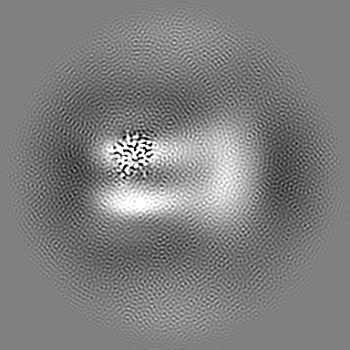
| Projections & Slices |
| ||||||||||||
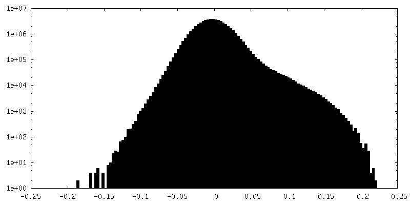
| Density Histograms |


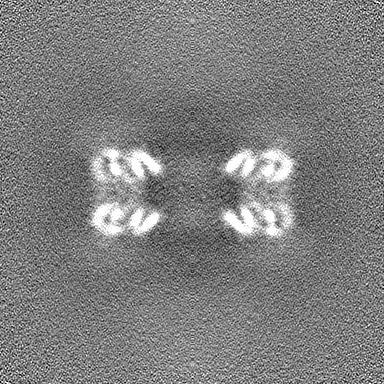
-Half map: CryoEM structure of ADP-DNA-MuB conformation2 with D10
| File | emd_38697_half_map_1.map | ||||||||||||
|---|---|---|---|---|---|---|---|---|---|---|---|---|---|
| Annotation | CryoEM structure of ADP-DNA-MuB conformation2 with D10 | ||||||||||||
| Projections & Slices |
| ||||||||||||
| Density Histograms |

-Half map: CryoEM structure of ADP-DNA-MuB conformation2 with D10
| File | emd_38697_half_map_2.map | ||||||||||||
|---|---|---|---|---|---|---|---|---|---|---|---|---|---|
| Annotation | CryoEM structure of ADP-DNA-MuB conformation2 with D10 | ||||||||||||
| Projections & Slices |
| ||||||||||||
| Density Histograms |


- Sample components
-Entire : ADP-DNA-MuB conformation2 with D10 in Bacteriophage Mu DNA Transp...
| Entire | Name: ADP-DNA-MuB conformation2 with D10 in Bacteriophage Mu DNA Transposition |
|---|---|
| Components |
|
-Supramolecule #1: ADP-DNA-MuB conformation2 with D10 in Bacteriophage Mu DNA Transp...
| Supramolecule | Name: ADP-DNA-MuB conformation2 with D10 in Bacteriophage Mu DNA Transposition type: complex / ID: 1 / Parent: 0 / Macromolecule list: #1 |
|---|---|
| Source (natural) | Organism: Escherichia phage Mu (virus) |
-Macromolecule #1: ATP-dependent target DNA activator B
| Macromolecule | Name: ATP-dependent target DNA activator B / type: protein_or_peptide / ID: 1 / Number of copies: 20 / Enantiomer: LEVO EC number: Hydrolases; Acting on acid anhydrides; In phosphorus-containing anhydrides |
|---|---|
| Source (natural) | Organism: Escherichia phage Mu (virus) |
| Molecular weight | Theoretical: 35.153133 KDa |
| Recombinant expression | Organism:  |
| Sequence | String: MNISDIRAGL RTLVENEETT FKQIALESGL STGTISSFIN DKYNGDNERV SQMLQRWLEK YHAVAELPEP PRFVETQTVK QIWTSMRFA SLTESIAVVC GNPGVGKTEA AREYRRTNNN VWMITITPSC ASVLECLTEL AFELGMNDAP RRKGPLSRAL R RRLEGTQG ...String: MNISDIRAGL RTLVENEETT FKQIALESGL STGTISSFIN DKYNGDNERV SQMLQRWLEK YHAVAELPEP PRFVETQTVK QIWTSMRFA SLTESIAVVC GNPGVGKTEA AREYRRTNNN VWMITITPSC ASVLECLTEL AFELGMNDAP RRKGPLSRAL R RRLEGTQG LVIIDEADHL GAEVLEELRL LQESTRIGLV LMGNHRVYSN MTGGNRTVEF ARLFSRIAKR TAINKTKKAD VK AIADAWQ INGEKELELL QQIAQKPGAL RILNHSLRLA AMTAHGKGER VNEDYLRQAF RELDLDVDIS TLLRN UniProtKB: ATP-dependent target DNA activator B |
-Macromolecule #2: ADENOSINE-5'-DIPHOSPHATE
| Macromolecule | Name: ADENOSINE-5'-DIPHOSPHATE / type: ligand / ID: 2 / Number of copies: 20 / Formula: ADP |
|---|---|
| Molecular weight | Theoretical: 427.201 Da |
| Chemical component information |  ChemComp-ADP: |
-Experimental details
-Structure determination
| Method | cryo EM |
|---|---|
 Processing Processing | single particle reconstruction |
| Aggregation state | particle |
-Sample preparation
| Buffer | pH: 7.8 |
|---|---|
| Vitrification | Cryogen name: ETHANE |
- Electron microscopy
| Microscope | FEI TITAN KRIOS |
|---|---|
| Image recording | Film or detector model: GATAN K3 (6k x 4k) / Number real images: 9510 / Average electron dose: 60.9 e/Å2 |
| Electron beam | Acceleration voltage: 300 kV / Electron source:  FIELD EMISSION GUN FIELD EMISSION GUN |
| Electron optics | Illumination mode: FLOOD BEAM / Imaging mode: BRIGHT FIELD / Nominal defocus max: 3.0 µm / Nominal defocus min: 2.0 µm |
| Experimental equipment |  Model: Titan Krios / Image courtesy: FEI Company |
