 Movie
Movie Controller


+ Open data

- Basic information
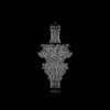
| Entry | Database: PDB / ID: 8vxq | |||||||||||||||||||||||||||||||||||||||||||||
|---|---|---|---|---|---|---|---|---|---|---|---|---|---|---|---|---|---|---|---|---|---|---|---|---|---|---|---|---|---|---|---|---|---|---|---|---|---|---|---|---|---|---|---|---|---|---|
| Title | Cryo-EM structure of phage DEV ejection proteins gp72:gp73 | |||||||||||||||||||||||||||||||||||||||||||||
 Components Components |
| |||||||||||||||||||||||||||||||||||||||||||||
 Keywords Keywords | VIRAL PROTEIN / phage / bacteriophage / STRUCTURAL PROTEIN / outer membrane protein / gp73 / gp74 / DEV | |||||||||||||||||||||||||||||||||||||||||||||
| Function / homology | Uncharacterized protein / N4 gp52-like protein Function and homology information Function and homology information | |||||||||||||||||||||||||||||||||||||||||||||
| Biological species |  Pseudomonas phage vB_PaeP_DEV (virus) Pseudomonas phage vB_PaeP_DEV (virus) | |||||||||||||||||||||||||||||||||||||||||||||
| Method | ELECTRON MICROSCOPY / single particle reconstruction / cryo EM / Resolution: 3.1 Å | |||||||||||||||||||||||||||||||||||||||||||||
 Authors Authors | Iglesias, S.M. / Cingolani, G. | |||||||||||||||||||||||||||||||||||||||||||||
| Funding support |  United States, 1items United States, 1items
| |||||||||||||||||||||||||||||||||||||||||||||
 Citation Citation | Journal: Nat Commun / Year: 2024 Title: Integrative structural analysis of Pseudomonas phage DEV reveals a genome ejection motor. Authors: Ravi K Lokareddy / Chun-Feng David Hou / Francesca Forti / Stephano M Iglesias / Fenglin Li / Mikhail Pavlenok / David S Horner / Michael Niederweis / Federica Briani / Gino Cingolani /  Abstract: DEV is an obligatory lytic Pseudomonas phage of the N4-like genus, recently reclassified as Schitoviridae. The DEV genome encodes 91 ORFs, including a 3398 amino acid virion-associated RNA polymerase ...DEV is an obligatory lytic Pseudomonas phage of the N4-like genus, recently reclassified as Schitoviridae. The DEV genome encodes 91 ORFs, including a 3398 amino acid virion-associated RNA polymerase (vRNAP). Here, we describe the complete architecture of DEV, determined using a combination of cryo-electron microscopy localized reconstruction, biochemical methods, and genetic knockouts. We built de novo structures of all capsid factors and tail components involved in host attachment. We demonstrate that DEV long tail fibers are essential for infection of Pseudomonas aeruginosa but dispensable for infecting mutants with a truncated lipopolysaccharide devoid of the O-antigen. We determine that DEV vRNAP is part of a three-gene operon conserved in 191 Schitoviridae genomes. We propose these three proteins are ejected into the host to form a genome ejection motor spanning the cell envelope. We posit that the design principles of the DEV ejection apparatus are conserved in all Schitoviridae. #1: Journal: Res Sq / Year: 2024 Title: Integrative structural analysis of phage DEV reveals a genome ejection motor. Authors: Gino Cingolani / Ravi Lokareddy / Chun-Feng Hou / Francesca Forti / Stephano Iglesias / Fenglin Li / Mikhail Pavlenok / Michael Niederweis / Federica Briani Abstract: DEV is an obligatory lytic phage of the N4-like genus, recently reclassified as . The DEV genome encodes 91 ORFs, including a 3,398 amino acid virion-associated RNA polymerase. Here, we describe the ...DEV is an obligatory lytic phage of the N4-like genus, recently reclassified as . The DEV genome encodes 91 ORFs, including a 3,398 amino acid virion-associated RNA polymerase. Here, we describe the complete architecture of DEV, determined using a combination of cryo-electron microscopy localized reconstruction, biochemical methods, and genetic knockouts. We built de structures of all capsid factors and tail components involved in host attachment. We demonstrate that DEV long tail fibers are essential for infection of and dispensable for infecting mutants with a truncated lipopolysaccharide devoid of the O-antigen. We identified DEV ejection proteins and, unexpectedly, found that the giant DEV RNA polymerase, the hallmark of the family, is an ejection protein. We propose that DEV ejection proteins form a genome ejection motor across the host cell envelope and that these structural principles are conserved in all . | |||||||||||||||||||||||||||||||||||||||||||||
| History |
|
- Structure visualization
| Structure viewer | Molecule: MolmilJmol/JSmol |
|---|
- Downloads & links
-Download
| PDBx/mmCIF format | 8vxq.cif.gz | 774.3 KB | Display | PDBx/mmCIF format |
|---|---|---|---|---|
| PDB format | pdb8vxq.ent.gz | 640.1 KB | Display | PDB format |
| PDBx/mmJSON format | 8vxq.json.gz | Tree view | PDBx/mmJSON format | |
| Others |  Other downloads Other downloads |
-Validation report
| Arichive directory | https://data.pdbj.org/pub/pdb/validation_reports/vx/8vxqftp://data.pdbj.org/pub/pdb/validation_reports/vx/8vxq | HTTPS FTP |
|---|
-Related structure data
| Related structure data |  43629MC 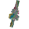 9bgmC 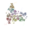 9bgnC  9bgoC  9codC M: map data used to model this data C: citing same article ( |
|---|---|
| Similar structure data |


-Links
 PDBj
PDBj- Assembly
| Deposited unit | 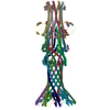
|
|---|---|
| 1 |
|
-Components
| #1: Protein | Mass: 57183.238 Da / Num. of mol.: 9 / Source method: isolated from a natural source / Source: (natural) Pseudomonas phage vB_PaeP_DEV (virus) / References: UniProt: A0A2K8HKQ8#2: Protein | Mass: 16639.414 Da / Num. of mol.: 9 / Source method: isolated from a natural source / Source: (natural) Pseudomonas phage vB_PaeP_DEV (virus) / References: UniProt: A0A2K8I0A4Has protein modification | N | |
|---|
-Experimental details
-Experiment
| Experiment | Method: ELECTRON MICROSCOPY |
|---|---|
| EM experiment | Aggregation state: PARTICLE / 3D reconstruction method: single particle reconstruction |
- Sample preparation
| Component | Name: Pseudomonas phage vB_PaeP_DEV / Type: VIRUS / Entity ID: all / Source: NATURAL |
|---|---|
| Molecular weight | Experimental value: NO |
| Source (natural) | Organism: Pseudomonas phage vB_PaeP_DEV (virus) |
| Details of virus | Empty: NO / Enveloped: NO / Isolate: OTHER / Type: VIRION |
| Natural host | Organism: Pseudomonas aeruginosa |
| Buffer solution | pH: 7.5 |
| Specimen | Embedding applied: NO / Shadowing applied: NO / Staining applied: NO / Vitrification applied: YES |
| Specimen support | Grid material: COPPER / Grid mesh size: 300 divisions/in. / Grid type: Quantifoil R2/1 |
| Vitrification | Instrument: FEI VITROBOT MARK IV / Cryogen name: ETHANE / Humidity: 100 % / Chamber temperature: 277.15 K |
- Electron microscopy imaging
| Experimental equipment |  Model: Titan Krios / Image courtesy: FEI Company |
|---|---|
| Microscopy | Model: FEI TITAN KRIOS |
| Electron gun | Electron source:  FIELD EMISSION GUN / Accelerating voltage: 300 kV / Illumination mode: OTHER FIELD EMISSION GUN / Accelerating voltage: 300 kV / Illumination mode: OTHER |
| Electron lens | Mode: OTHER / Nominal defocus max: 2200 nm / Nominal defocus min: 800 nm / Cs: 2.7 mm / C2 aperture diameter: 100 µm |
| Specimen holder | Cryogen: NITROGEN / Specimen holder model: FEI TITAN KRIOS AUTOGRID HOLDER |
| Image recording | Electron dose: 1 e/Å2 / Film or detector model: FEI FALCON IV (4k x 4k) |
- Processing
| EM software | Name: PHENIX / Category: model refinement | ||||||||||||||||||||||||
|---|---|---|---|---|---|---|---|---|---|---|---|---|---|---|---|---|---|---|---|---|---|---|---|---|---|
| CTF correction | Type: PHASE FLIPPING AND AMPLITUDE CORRECTION | ||||||||||||||||||||||||
| Symmetry | Point symmetry: C9 (9 fold cyclic) | ||||||||||||||||||||||||
| 3D reconstruction | Resolution: 3.1 Å / Resolution method: FSC 0.143 CUT-OFF / Num. of particles: 61482 / Algorithm: FOURIER SPACE / Num. of class averages: 1 / Symmetry type: POINT | ||||||||||||||||||||||||
| Refine LS restraints |
|
