National Institutes of Health/National Institute of General Medical Sciences (NIH/NIGMS)
135343
米国
National Institutes of Health/Eunice Kennedy Shriver National Institute of Child Health & Human Development (NIH/NICHD)
110229
米国
Welch Foundation
I-1957
米国
引用
ジャーナル: Nat Commun / 年: 2024 タイトル: Structure and inhibition of the human lysosomal transporter Sialin. 著者: Philip Schmiege / Linda Donnelly / Nadia Elghobashi-Meinhardt / Chia-Hsueh Lee / Xiaochun Li / 要旨: Sialin, a member of the solute carrier 17 (SLC17) transporter family, is unique in its ability to transport not only sialic acid using a pH-driven mechanism, but also transport mono and diacidic ...Sialin, a member of the solute carrier 17 (SLC17) transporter family, is unique in its ability to transport not only sialic acid using a pH-driven mechanism, but also transport mono and diacidic neurotransmitters, such as glutamate and N-acetylaspartylglutamate (NAAG), into synaptic vesicles via a membrane potential-driven mechanism. While most transporters utilize one of these mechanisms, the structural basis of how Sialin transports substrates using both remains unclear. Here, we present the cryogenic electron-microscopy structures of human Sialin: apo cytosol-open, apo lumen-open, NAAG-bound, and inhibitor-bound. Our structures show that a positively charged cytosol-open vestibule accommodates either NAAG or the Sialin inhibitor Fmoc-Leu-OH, while its luminal cavity potentially binds sialic acid. Moreover, functional analyses along with molecular dynamics simulations identify key residues in binding sialic acid and NAAG. Thus, our findings uncover the essential conformational states in NAAG and sialic acid transport, demonstrating a working model of SLC17 transporters.
 ムービー
ムービー コントローラー


 データを開く
 基本情報
基本情報 要素
要素 キーワード
キーワード 機能・相同性情報
 Homo sapiens (ヒト)
 データ登録者
 米国, 3件
  引用
引用
 構造の表示
構造の表示 ダウンロードとリンク
 その他のダウンロード


 PDBj
PDBj


 集合体
集合体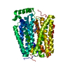

 分子量: 44.053 Da / 分子数: 1 / 由来タイプ: 合成 / 式: C2H4O / タイプ: SUBJECT OF INVESTIGATION
 タイプ: L-peptide linking / 分子量: 133.103 Da / 分子数: 1 / 由来タイプ: 合成 / 式: C4H7NO4 / タイプ: SUBJECT OF INVESTIGATION
 タイプ: L-peptide linking / 分子量: 147.129 Da / 分子数: 1 / 由来タイプ: 合成 / 式: C5H9NO4 / タイプ: SUBJECT OF INVESTIGATION
 試料調製
試料調製 電子顕微鏡撮影
 FIELD EMISSION GUN / 加速電圧: 300 kV / 照射モード: FLOOD BEAM
 解析
解析